


Abstract
Insomnia is the most prevalent sleep disorder worldwide, affecting up to 30% of the population, and is frequently accompanied by cognitive complaints and measurable cognitive impairments. Chronic insomnia is characterized by persistent hyperarousal across physiological, cognitive, and neurobiological domains, which may compromise multiple cognitive systems. This review synthesizes current evidence on the impact of insomnia on major cognitive domains, including attention, language, memory and learning, executive functions, perceptual–motor skills, and social cognition. We critically evaluate methodological factors contributing to heterogeneous findings across studies, such as variability in diagnostic criteria, insomnia phenotypes, cognitive assessments, and sample characteristics. Additionally, we summarize emerging evidence on neuroanatomical and molecular correlates of insomnia-related cognitive impairment, including alterations in hippocampal and prefrontal brain structures, amyloid-β accumulation, dysregulation of brain-derived neurotrophic factor, neurotransmitter imbalance, neuroinflammation, and disrupted signaling pathways. Insights from preclinical studies show mechanistic links between sleep loss, synaptic dysfunction, oxidative stress, and memory decline. Despite substantial advances, the precise pathophysiological mechanisms underlying cognitive dysfunction in insomnia remain incompletely understood, suggesting that cognitive impairment arises from the convergence of multiple biological processes rather than a single causal pathway. A deeper mechanistic understanding is essential for developing targeted interventions for preventing cognitive decline in individuals with insomnia.
1. Introduction
Insomnia represents one of the most prevalent sleep disorders, affecting more than 16% of the global population [1]. Recent epidemiological reports showed that ~10% of adults actually suffer from an insomnia disorder, while another ~20% experience occasional insomnia symptoms, meaning that approximately 30% of the general population have some insomnia symptoms during their lifetime [2]. Importantly, 40–50% of adults with insomnia have coexisting comorbid psychiatric conditions, especially anxiety and depression [3].
Insomnia was first described by Johann Heinroth in 1818 as a state characterized by inadequate sleep quality and quantity [4]. Traditionally considered as a subjective complaint, insomnia is commonly assessed by asking patients whether they have trouble falling or staying asleep. However, it can also be objectively characterized using polysomnography. Therefore, insomnia may be considered as both a symptom and a clinical sign [5,6]. According to the Fifth Edition of the Diagnostic and Statistical Manual for Mental Disorders (DSM-5), insomnia disorder is defined by difficulty initiating or maintaining sleep, or early morning awakening with an inability to return to sleep, occurring at least three times per week for a minimum period of three months. These symptoms must persist despite adequate sleep opportunity and cannot be better explained by another medical, psychiatric, or sleep disorder, nor by substance use [7].
Historically, “primary insomnia (PI)” referred to cases without identifiable medical or psychiatric causes. However, contemporary classifications such as DSM-5 and the International Classification of Sleep Disorders Third Edition (ICSD-3) have largely replaced this concept with the broader term insomnia disorder [7,8]. Additionally, the term “comorbid insomnia” has replaced the outdated term “secondary insomnia”, recognizing that coexisting mental or somatic conditions may contribute to sleep disturbances without diminishing the importance of treating insomnia itself.
Beyond its well-established impact on mood and quality of life [9], growing evidence suggests that insomnia is associated with impairments across multiple cognitive domains [10]. Definitions of cognitive performance differ considerably across studies, but are most often operationalized as either subjective measures (based on self-reports) or objective measures (based on cognitive tasks or tests) [11]. Emerging evidence suggests a bidirectional relationship between insomnia and cognitive dysfunction: sleep disturbances can exacerbate cognitive impairment, while cognitive deficits may further perpetuate sleep difficulties [12].
Neurobiological models propose that alterations in prefrontal cortex functioning, dysregulation of the hypothalamic–pituitary–adrenal (HPA) axis, and impaired synaptic plasticity may underlie these cognitive deficits [13]. These mechanisms highlight that insomnia is not merely a symptom but also a potential contributor to long-term neurocognitive vulnerability. However, although patients with insomnia frequently report cognitive complaints, and experimental sleep deprivation (SD) shows clear impairments in several cognitive functions, these findings cannot be directly generalized to insomnia, as it is not necessarily defined by objective SD [14]. Despite increasing research interest, findings remain heterogeneous, likely due to differences in diagnostic criteria, insomnia phenotypes, cognitive assessments, and neurobiological measures.
This article represents a narrative review of clinical and preclinical studies addressing the relationship between insomnia and cognitive functions. Additionally, we integrated structural, molecular, and mechanistic evidence from both human and animal studies, providing a framework for understanding how sleep disruption leads to cognitive impairment.
2. Methods of Review
This manuscript represents a narrative review of clinical and preclinical studies, which examined the relationship between insomnia and cognitive dysfunction, with particular emphasis on neurobiological mechanisms. A literature search was conducted using PubMed/MEDLINE, Scopus, and Web of Science databases. Searches included articles published up to February 2026, with emphasis on studies from the past 15 years to reflect contemporary diagnostic criteria and neurobiological advances. Key search terms were combined using Boolean operators and included: “insomnia”, “insomnia disorder”, “chronic insomnia”, “sleep deprivation”, “cognition”, “cognitive functions”, “attention”, “memory”, “language”, “visual-motor function”, “executive function”, “social cognition”, “cognitive impairment”, “cognitive dysfunction”, “neuroimaging”, “neurotransmitters”, “hippocampus”, “prefrontal cortex”, “amyloid-β”, “BDNF”, “neuroinflammation”, “oxidative stress”, and “signaling pathways”.
Inclusion criteria comprised: (1) peer-reviewed original clinical studies, (2) longitudinal or cross-sectional human investigations assessing cognitive performance in insomnia, (3) neuroimaging or biomarker studies exploring structural, functional, or molecular correlates, and (4) preclinical studies employing validated sleep deprivation paradigms that provided mechanistic insight relevant to cognitive outcomes. Systematic reviews and meta-analyses were prioritized when available. Exclusion criteria included: (1) case reports, (2) studies focusing exclusively on other primary sleep disorders (e.g., obstructive sleep apnea or narcolepsy) without insomnia-specific data, and (3) studies lacking objective or clearly defined cognitive measures.
Given the methodological heterogeneity across studies—including differences in diagnostic criteria, insomnia phenotypes (subjective vs. objectively short sleep duration), cognitive assessment tools, and study design—findings were synthesized narratively. Greater interpretative weight was given to studies with larger samples, objective sleep measures, standardized neuropsychological testing, and longitudinal designs. When conflicting results were identified, potential sources of discrepancy were critically examined.
3. Mechanisms of Insomnia in Brief
There is a growing understanding that insomnia often develops through complex interactions among environmental, behavioral, and biological factors rather than a simple linear cause (Figure 1). Given the distinct clinical courses and potentially unique pathophysiological mechanisms and specific insomnia background, differentiation remains important for selecting appropriate therapeutic strategies [15]. Insomnia has been conceptualized as a disorder of heightened psychological and physiological arousal, characterized by elevated alertness during the day and difficulty initiating or maintaining sleep at night [16]. Several mechanistic models have been proposed to explain the development and chronicity of insomnia. The common feature of these models is the central role of heightened arousal across cognitive–emotional, cortical, and/or physiological domains [17].
Figure 1.
The interplay between environmental, behavioral, and biological factors in the development of insomnia. Abbreviations: GABA—gamma-aminobutyric acid; HPA—hypothalamic–pituitary–adrenal.
The physiological model suggests that chronic activation of neurophysiological systems contributes to a persistent hyperarousal state. In patients with insomnia, evidence of physiological arousal has been demonstrated in insomnia patients via various metabolic rate measurements, heart rate variability, neuroendocrine measurements, and functional neuroimaging [18]. Metabolic rate elevation can be estimated through oxygen consumption levels [19], while markers such as elevated evening cortisol, increased heart rate, changes in autonomic balance, heightened metabolism, and enhanced electroencephalographic (EEG) activity (16–50 Hz) reflect this arousal pattern [20]. Importantly, these physiological disturbances may interfere with sleep initiation and maintenance, supporting the hypothesis that insomnia represents a disorder of chronic hyperactivation.
The cognitive model emphasizes that worry, rumination, and excessive focus on sleep and its consequences play a central role in perpetuating insomnia [21]. Stressful life events may trigger acute episodes of sleep disturbance, which can evolve into chronic insomnia when maladaptive cognitive patterns persist. This model highlights the role of dysfunctional beliefs about sleep—such as catastrophic interpretations of fatigue—in maintaining the disorder.
The behavioral model, besides the sleep hygiene and stimulus–control model, emphasize Spielman’s model (3P-model), which identifies the interplay of predisposing, precipitating, and perpetuating factors as a trigger for increased insomnia burden [22,23]. Factors that predispose individuals to develop insomnia include genetic vulnerability, personality characteristics such as neuroticism or perfectionism, a tendency to inhibit negative emotions, and an increased susceptibility to both cognitive and physiological hyperarousal [24]. Even though perpetuating factors represent a myriad of compensatory strategies initially developed for sleep improvement (such as early bedtime schedule), these patterns may also induce a mismatch between opportunity and the real ability for sleeping, further enhancing insomnia (Figure 2) [25]. Recognition of perpetuating factors is essential in clinical practice, forming the basis of cognitive behavioral therapy for insomnia (CBT-I), the first-line treatment [26].
Figure 2.
Visual representation of Spielman’s model (3P-model). Insomnia severity changes over time as a function of predisposing, precipitating, and perpetuating factors. Predisposing factors create a baseline vulnerability that typically remains below the insomnia threshold. Acute precipitating factors trigger a rise in insomnia severity above this threshold, initiating insomnia. Over time, perpetuating factors maintain elevated insomnia severity even after the initial trigger resolves, contributing to the development and persistence of chronic insomnia.
Finally, the neurocognitive model observes arousal across three pathways—somatic, cognitive, and cortical, with cortical arousal reflected by the detection of high-frequency EEG activity during sleep [25]. Such elevated activity is thought to reflect enhanced sensory processing, memory-related activity, and heightened conscious perception during sleep [27]. This model bridges physiological and psychological frameworks by highlighting how neural hyperactivation translates into subjective experiences of light, unrefreshing sleep.
4. Insomnia and Main Cognitive Domains
The word cognition originates from the Latin word “cognoscere”, which means “to know with”, related with “to understand” [28]. Each newly generated piece of information involves the activity of cognitive functions in an already adjusted common order. Therefore, when our sensory system receives new information, it is then chosen for further processing via attention and can be retained during a short period by the working memory system, while the long-term memory is associated with language functions [29]. According to DSM-5, the entire cognitive system refers to six main domains—attention, learning and memory, language, executive functions, perceptual–motor functions (with visuospatial function), and social cognition functions—each divided into specific subdomains. On the other hand, cognitive disorders include delirium, mild neurocognitive disorder, and major neurocognitive disorder—dementia [7]. As cognition itself contains a voluntary component and influences emotional reactions and decision-making in every individual, there are no clear boundaries between cognitive, emotional, and voluntary functions [29]. Sufficient sleep is crucial for preserving attentional control, cognitive alertness, and sensory processing, thereby supporting optimal performance in tasks that demand sustained focus [30].
Despite the well-established role of sleep in cognitive functioning, the literature examining cognitive performance in insomnia remains heterogeneous. Some studies report measurable impairments across several domains, whereas others find minimal or no objective deficits. Importantly, these inconsistencies appear to reflect not only genuine variability in cognitive functioning among individuals with insomnia but also substantial differences in study design, sample characteristics, and cognitive assessment methods. When the literature is considered collectively, a pattern emerges suggesting that impairments tend to be subtle and domain-specific rather than representing a global cognitive deficit.
Attention is a state of mental orientation which involves focusing on central components in our consciousness while neglecting all irrelevant components [31], and can be examined through subdomains such as attention span, focused attention, divided attention, sustained attention, and other complex attention tasks.
Evidence generally suggests that basic attentional processes are relatively preserved in insomnia. Several studies report preserved performance in basic attentional processes such as sustained [32,33,34,35] and focused [35,36,37,38,39] attention. However, other investigations have observed poorer sustained attention in individuals with insomnia compared to healthy sleepers [39,40]. These inconsistencies may partly reflect methodological differences across studies, particularly the complexity of the cognitive tasks employed. Tasks assessing simple reaction time or basic vigilance may lack the sensitivity to detect subtle impairments, whereas those requiring more complex attentional processing appear more likely to reveal deficits. Consistent with this interpretation, Fortier-Brochu and colleagues reported mild-to-moderate impairment in several attention processes in insomnia patients, including longer response and information-processing times, followed by poorer outcomes for selective attention. However, other aspects of attention, such as alertness, shared attention, and sustained attention, did not differ significantly between insomnia patients and healthy sleepers [41,42]. Meta-analytic evidence largely aligns with this pattern, suggesting that cognitive deficits in insomnia are selective rather than generalized [11]. Specifically, insomnia has been linked to objective impairments in alertness, complex attention, working memory (retention and manipulation), episodic memory, executive function, and perceptual processing. In contrast, psychomotor skills, verbal abilities, information processing, various forms of attention (selective, divided, sustained), procedural memory, verbal fluency, and cognitive flexibility appear largely unaffected [11]. Moreover, insomnia without excessive sleepiness was associated with greater attentional impairment, which may negatively affect work productivity [43].
Evidence regarding more complex attentional functions is particularly mixed. Studies examining attention-shifting—the most demanding attentional process—have produced inconsistent findings. Some reports indicate greater deficits in attention shifting among insomnia patients [37,44], whereas others find no significant differences compared to healthy sleepers [45,46]. Nevertheless, impairments in set-switching attention [47], and higher-order attentional functions [48] have been observed in patients with chronic insomnia and objectively short sleep duration, suggesting that attentional deficits may be more pronounced in specific insomnia phenotypes.
For other attentional domains, including attention span, divided attention, and complex focused attention, most studies report no significant differences between patients with insomnia and controls [33,49,50,51,52,53,54]. However, some evidence indicates deficits in specific task components, such as forward and reversed digit span [33,39], perceptual speed [49], detectability, and perseverative errors [42], and reduced vigilance performance in at least one study involving insomnia patients [50].
These discrepancies likely reflect heterogeneity in task selection, outcome measures, and sample characteristics. In particular, differences in task complexity may affect whether subtle attentional deficits are detectable. Basic reaction-time tasks often fail to detect impairments, whereas more complex tasks that require attentional switching or high cognitive load are more likely to reveal deficits. Supporting these interpretations, evidence from a large cohort of 1000 adults showed that subjective attention complaints in insomnia were not corroborated by objective testing [55]. Taken together, these findings suggest that attentional deficits in insomnia may reflect reduced efficiency in higher-order attentional control processes rather than a generalized impairment of basic attentional capacity. Collectively, these findings underscore the need for carefully standardized methodologies to clarify the impact of insomnia on attentional performance.
Language, a process of articulating meaningful speech, relies on multiple cognitive abilities, including word and sentence analysis [56]. Although sleep disruption has the potential to influence these processes, evidence regarding language functioning in insomnia remains limited and somewhat inconsistent. Experimental sleep deprivation studies suggest that acute sleep loss may reduce verbal fluency [57,58] and other linguistic skills [59], potentially by limiting word generation and the reliance on semantic networks. However, these findings are not consistently replicated. Some investigations report reduced verbal fluency following a sleepless night, whereas others observe no decline or even modest improvements in certain verbal tasks [60]. Such variability may reflect differences in task demands, motivational factors, or compensatory cognitive strategies during short-term sleep loss. Interestingly, individuals exposed to SD performed worse when following instructions but better when giving instructions in model-building tasks [61].
In contrast, studies examining sleep disturbances, specifically insomnia, generally indicate that language abilities are relatively preserved compared with other cognitive domains [62]. These mixed findings imply that the relationship between insomnia and language is non-linear and context-dependent, influenced by factors such as task complexity, the chronicity of sleep disturbance, individual cognitive reserve, and motivation. Limitations of existing research—including small samples, simplistic tasks, and cross-sectional designs—further complicate interpretation. Taken together, current evidence suggests that language abilities are relatively resilient to sleep disturbance, although subtle inefficiencies may emerge under conditions of acute sleep loss or increased linguistic demand. Future studies using mechanistic, fine-grained approaches that integrate behavioral, neurocognitive, and linguistic measures are needed to clarify how different aspects of sleep disruption affect specific components of language production and comprehension.
Memory represents a mental function responsible for remembering or reproducing the content that is already a part of an experience or has already been learned. On the other hand, learning enables the acquisition of new or modification of existing knowledge, behavior, skills, values, or preferences [63]. To investigate cognitive differences between individuals with insomnia and healthy sleepers, studies commonly assess working, implicit, and explicit memory domains [11].
Overall, the literature suggests that insomnia may be associated with impairments in several memory processes, although findings are not entirely consistent. Evidence most frequently points to deficits in episodic memory, with Fortier-Brochu and colleagues reporting poorer episodic memory performance in insomnia patients compared with controls [42]. Additional studies have also reported deficits in related domains, including immediate memory, delayed recall, emotional memory, and decision-making among individuals with insomnia [64,65]. However, despite substantial evidence linking insomnia to memory dysfunction, some investigations have failed to detect widespread memory impairments. Instead, these studies report group differences limited to specific domains, such as perceptual organization, which involves non-verbal reasoning and visuospatial integration [66].
These discrepancies may partly reflect methodological limitations, including small sample sizes and variability in cognitive paradigms. Nevertheless, findings from larger population-based studies provide stronger support for an association between insomnia and cognitive decline. For example, evidence from a large Canadian cohort of nearly 30,000 adults aged 45 years and older demonstrated that chronic insomnia is independently associated with poorer cognitive functioning, regardless of comorbid conditions. Notably, declarative memory appears to be particularly vulnerable, with PI patients showing greater impairment compared with individuals without insomnia symptoms or those with insomnia symptoms but preserved daytime functioning [67].
Neuroimaging studies further support the presence of memory-related neural dysfunction in insomnia. Using functional magnetic resonance imaging (MRI), Drummond and colleagues reported that patients with PI exhibited reduced activation of brain regions typically engaged during working memory tasks. Unlike good sleepers, insomnia patients showed neither compensatory recruitment of alternative brain areas nor task-dependent modulation of neural activity during increasing cognitive demands [68]. These findings suggest altered neural efficiency and impaired functional adaptability, providing neurophysiological evidence that complements behavioral observations of memory deficits in insomnia. Collectively, these findings may indicate that insomnia selectively affects memory processes that depend on efficient hippocampal and prefrontal functioning.
Executive functions encompass an integrated composite of many related elemental cognitive activities, such as motivation, making plans, thinking ahead, suppressing irrelevant sensory inputs, imagining, evaluating, changing or correcting behavior, and control in initiating goal-oriented basic and complex actions, as well as more basic executive processes (shifting, updating and inhibition) [69]. As higher mental functions, executive functions control the most complex forms of human activities, which are essential for achieving goals and enabling an individual to behave and respond adaptively in different situations [70].
Insomnia affects synapses, causing neural connections to slow down and consequently poor functioning, which can directly affect the performance of cognitive processes and executive functions [71]. Available evidence regarding the impact of sleep disruption on executive functions differs from study to study, indicating that scientific knowledge in this area is still evolving. For instance, some studies have reported small-to-moderate deficits in insomnia patients compared to controls while performing executive function tasks [41,42,44,47,72,73,74]; others reported no significant differences between these two groups [45,74,75,76,77]. Despite these inconsistencies, certain executive domains appear more consistently affected. In particular, impairments in decision-making, problem-solving, and the organization of ideas have frequently been reported in association with sleep disruption [11,78,79,80,81]. Evidence also suggests that executive functioning may be sensitive even to relatively short periods of sleep loss. For example, impairments in executive processes have been reported after 24 h of sleep restriction [82]. Furthermore, young PI patients have also exerted difficulties in working memory during high-demanding memory performance [83].
Overall, executive dysfunction in insomnia appears modest but may become more evident during tasks requiring complex cognitive control or sustained mental effort. Further research should be focused on determining the influence of insomnia on particular components of executive functions.
The visual–motor function involves the performance of different tasks (such as organization and manual manipulation with spatial information) along with skills including perceptual and visuospatial skills, programming and coordination as well as executive functioning [84]. The development of perceptual–motor functions includes the ability for spatial, directional, body, and temporal awareness. Compared with other cognitive domains, the relationship between insomnia and perceptual–motor functioning has been less extensively investigated. Existing evidence suggests that impairments in this domain may be relatively subtle. For example, one meta-analysis revealed small impairments in perceptual functioning in insomnia patients assessed for different tasks such as perceptual reasoning index and critical flicker functions [11]. Interestingly, it has been shown that in PI patients, the education level was markedly correlated with visuospatial function [64]. Nevertheless, to our knowledge, only a few mentioned studies investigated the impairment of perceptional functions in patients with insomnia, so any further conclusions regarding this topic would be unjustified.
Social cognition is based on cognitive processes that serve to establish and maintain social interactions (understanding the desires and needs of others) [85]. On the other hand, metacognition is also associated with social cognition, and represents the ability for deliberation of one’s own cognitive processes [86]. Facial recognition is one of the components of social cognition and reflects the ability to recognize the different emotional facial expressions.
Evidence examining social cognition in insomnia remains limited but suggests possible impairments in emotional processing. De Almondes and colleagues demonstrated a lower ability for recognizing facial emotions (fear and sadness) and decreased perceptual organization in insomnia patients compared to controls [66]. Furthermore, insomnia patients also performed worse during neuropsychological testing, which involved inhibitory control, planning capacity, problem-solving, and cognitive flexibility [66]. The possible explanation for these findings could be the impaired frontal–amygdala route in insomnia patients, which can compromise some emotional aspects involved in the recognition of fear [66]. Moreover, insomnia patients recognized angry facial expressions to a lesser extent than controls, due to impaired visual attention probably driven by emotional perception decline [87]. Most recently, one meta-analysis reported nearly 30% higher risk in insomnia patients for developing cognitive disorders [88], indicating that despite evident inconsistency between studies, further efforts are needed to resolve misrecognized areas in this neuropsychiatric field to improve the therapeutic approach in insomnia patients and prevent possible cognitive deterioration.
Potential Sources of Heterogeneity Across Studies
Across cognitive domains, inconsistent findings likely reflect several identifiable sources of methodological and clinical heterogeneity rather than the absence of true cognitive effects. Three major factors warrant consideration: (1) differentiation between acute sleep deprivation and chronic insomnia disorder, (2) variability in insomnia phenotyping (subjective versus objective sleep measures), and (3) differences in cognitive assessment methodology and study design.
First, acute sleep deprivation (SD) differs fundamentally from chronic insomnia. While chronic insomnia disorder involves ≥3 nights per week for ≥3 months [7], acute SD typically involves total sleep deprivation (e.g., 24 h or more of continuous wakefulness) or partial sleep restriction across one or two nights in otherwise healthy individuals [89]. Therefore, findings from SD studies cannot be directly extrapolated to insomnia disorder. SD typically produces robust cognitive impairments, with the acute SD followed by severe deficits. Interestingly, chronic partial sleep restriction tends to produce more subtle but cumulative cognitive deficits [90]. Cognitive impairments in chronic insomnia tend to be of a small-to-moderate magnitude [41]. Studies that do not clearly distinguish between these conditions may contribute to inconsistencies in the literature.
Second, heterogeneity arises from differences in insomnia definition and phenotyping. Some studies rely exclusively on subjective self-report measures such as the Insomnia Severity Index (ISI) or Pittsburgh Sleep Quality Index (PSQI), whereas others incorporate objective assessments including polysomnography or actigraphy. Emerging evidence suggests that insomnia accompanied by objectively short sleep duration represents a biologically more severe phenotype, associated with greater cognitive and neurobiological alterations, including dysregulation of the HPA axis, cardiometabolic risk, and impaired neurocognitive performance [91,92,93]. In contrast, insomnia with normal objective sleep duration may reflect heightened sleep misperception and cognitive–emotional hyperarousal rather than measurable cognitive inefficiency, and has been linked more strongly to psychological and behavioral factors [20,91]. Studies failing to distinguish these phenotypes may obscure subgroup-specific effects.
Third, there is considerable variability in the methods used to assess cognitive function. Different studies use various neuropsychological tests, computerized tasks, and experimental paradigms that measure related but not identical cognitive abilities. Differences in task sensitivity, cognitive load, and outcome measures may affect whether subtle impairments are detected. For instance, simple reaction-time or low-demand tasks often show normal performance, while more complex or demanding tasks are more likely to reveal cognitive impairment. In addition, studies with small samples and cross-sectional designs may lack sufficient power to detect small effects, whereas larger cohort studies often report more consistent associations.
Other factors may further complicate interpretation, including inconsistent control of psychiatric comorbidities, medication status, age-related cognitive vulnerability, and educational attainment. Importantly, many studies reporting null findings involve modest sample sizes or rely exclusively on subjective sleep measures, whereas studies demonstrating cognitive differences more frequently employ objective sleep assessment, standardized neuropsychological batteries, and larger samples.
When greater interpretative weight is given to methodologically rigorous studies, a clearer pattern emerges. These studies suggest selective vulnerability of attention, working memory, and executive functions [41], particularly under conditions of high cognitive demand or objectively short sleep duration.
Collectively, these sources of heterogeneity suggest that cognitive impairment in insomnia is neither universal nor absent; rather, it is conditional, phenotype-dependent, and sensitive to methodological design. A more precise characterization of insomnia subtypes and standardized cognitive assessment protocols will be essential for clarifying the true magnitude and clinical relevance of insomnia-related cognitive vulnerability.
For better understanding, various clinical and experimental studies investigating the relationship between sleep loss and cognitive impairment have provided significant evidence on structural and molecular mechanisms following worse cognitive outcomes in the setting of restricted sleep. Hence, in the next section we will summarize the current neurobiological findings on this topic.
5. Cognitive Impairment in Insomnia Patients—Neuroanatomical and Molecular Aspects
Chronic insomnia is increasingly recognized as a disorder extending beyond nighttime symptoms and daytime fatigue. Recent clinical studies have increasingly focused on the mechanistic basis of cognitive impairment associated with insomnia, examining structural, functional, and molecular abnormalities in the human brain [94]. Key findings are summarized in Table 1 and Table 2.
Table 1.
Neuroimaging findings associated with cognitive impairment in insomnia.
Table 2.
Molecular and inflammatory biomarkers associated with insomnia and cognitive impairment.
5.1. Brief Insights into Brain Morphology of Insomnia Patients
Advances in structural and functional neuroimaging have substantially improved our understanding of brain morphology alterations associated with insomnia. However, findings across studies remain heterogeneous, reflecting differences in diagnostic criteria, sample characteristics, insomnia phenotypes, and imaging methodologies. Among the most consistently investigated brain regions, the hippocampus has received particular attention due to its central role in memory processing and vulnerability to sleep disruption.
Early neuroimaging evidence demonstrated bilateral hippocampal volume reduction in PI patients compared with healthy controls, suggesting that chronic sleep disturbance may be associated with hippocampal structural vulnerability [95]. Subsequent studies yielded mixed results, with some reporting preserved whole-brain and hippocampal volumes in insomnia patients [96,100], while others identified more localized or subregional hippocampal abnormalities. For example, Joo and colleagues reported reduced hippocampal volume accompanied by atrophy across CA1–CA4 and dentate gyrus subfields, which was associated with impairments in memory, working memory, and verbal fluency [102]. Similarly, reduced hippocampal fimbria volume was observed in insomnia patients and correlated with greater symptom severity and higher depression and anxiety scores [105]. In patients with comorbid posttraumatic stress disorder, reductions in CA3/dentate gyrus subfields were reported, with hippocampal volume negatively correlating with insomnia severity, highlighting the sensitivity of hippocampal subregions to sleep disruption and stress-related arousal [99].
Beyond the hippocampus, alterations in additional brain regions involved in cognitive and emotional regulation have been described. Cortical thinning in the anterior cingulate cortex and shape deformation of the caudate nucleus were reported in a recent large structural MRI study, suggesting disruptions in cortico-striatal networks implicated in executive control and emotional processing [97]. Reductions in thalamic volume and decreased resting-state functional connectivity between the thalamus and frontal and limbic regions have also been identified, supporting impaired thalamo-cortical communication as a potential mechanism underlying attentional and cognitive complaints in insomnia [101].
Voxel-based morphometry studies further support region-specific gray matter alterations. Reductions in gray matter volume have been identified in the orbitofrontal cortex, parietal cortex, and the precuneus, regions implicated in executive functioning, self-referential processing, and attentional control [103]. Although some investigations found no global changes in gray or white matter volumes when normalized to total intracranial volume [96,107], others reported selective hippocampal volume loss in anterior subregions, suggesting that subtle, regionally confined alterations may not be captured by global volumetric measures [107].
Alterations in limbic structures beyond the hippocampus have also been reported. While some studies observed preserved amygdala volume overall, localized amygdala atrophy and reduced gray matter volume were identified in patients with chronic insomnia, indicating possible disruptions in emotional regulation and stress responsivity [104]. Functional adaptations have additionally been described, with increased resting-state functional connectivity between the hippocampal CA1 subregion and prefrontal cortex in certain insomnia phenotypes, potentially reflecting compensatory neural mechanisms [106]. The prefrontal cortex, as a crucial part of the cerebral cortex involved in performing executive functions, seems to be particularly vulnerable to prolonged wakefulness, especially with aging [121].
At the level of white matter integrity, large-scale population-based data suggest a potential link between insomnia and cerebrovascular vulnerability. Specifically, insomnia accompanied by short sleep duration has been associated with an increased burden of white matter hyperintensities, whereas longer sleep duration appears to exert a protective effect [108].
Taken together, neuroimaging findings indicate that insomnia is associated with subtle but meaningful alterations in brain morphology and connectivity, particularly within hippocampal, thalamic, prefrontal, and limbic networks. The variability of reported results likely reflects both methodological differences and genuine heterogeneity in insomnia presentations. These structural and functional alterations may represent important neurobiological substrates underlying cognitive impairment in insomnia, warranting further investigation using standardized diagnostic criteria, longitudinal designs, and high-resolution imaging approaches.
5.2. The Impact of Amyloid β (Aβ) Deposits
Insomnia and sleep disruption seem to be associated with alterations in amyloid-β (Aβ) metabolism [109], a hallmark of neurodegenerative processes and cognitive decline [122]. Both clinical and experimental studies indicate that impaired sleep continuity may interfere with Aβ clearance, thereby promoting its accumulation in vulnerable brain regions [112,113].
A bidirectional relationship between sleep disruption and Aβ accumulation has been suggested, with evidence derived from both human observational studies and experimental models. Aβ accumulation initiates sleep disruption, which in turn further promotes Aβ deposition [123]. The increase in Aβ deposition during sleep disruption has been hypothesized to involve dysregulation of the glymphatic system, a cerebrospinal fluid clearance pathway [124].
Evidence from CSF studies supports the presence of early amyloid dysregulation in insomnia. Investigations in PI patients have demonstrated increased concentrations of Aβ species. Cheng and colleagues reported elevated CSF Aβ42 and Aβ40 levels in insomnia patients compared with healthy controls, while total Tau protein (Tau) and phosphorylated Tau (p-Tau) levels remained unchanged [109], suggesting early amyloid dysregulation in the absence of overt neurodegeneration. Similarly, higher insomnia severity, indexed by the ISI, was associated with increased CSF Aβ42 concentrations, whereas Tau and neurofilament light chain (NfL) levels were not significantly altered, which may suggest that Aβ changes precede downstream neurodegenerative markers [111].
Neuroimaging studies using positron emission tomography (PET) have provided complementary evidence of amyloid burden in insomnia. In a large clinical cohort, insomnia patients exhibited increased baseline amyloid PET burden compared with controls, although longitudinal amyloid accumulation did not differ significantly over time, indicating that insomnia may influence early amyloid deposition rather than accelerating progressive accumulation [108]. Experimental SD paradigms reinforce this interpretation. Experimental sleep deprivation studies in humans have shown transient increases in Aβ levels in regions such as the hippocampus, parahippocampal gyrus, and thalamus, regions critically involved in memory consolidation and arousal regulation [112]. In older adults, disrupted sleep was associated with greater hippocampal Aβ accumulation and impaired episodic associative memory, underscoring the cognitive relevance of sleep-dependent amyloid regulation [110].
Importantly, sleep architecture appears to play a pivotal role in Aβ dynamics. Reduced slow-wave activity, a defining feature of fragmented and insufficient sleep, has been linked to increased plasma Aβ40/42 ratios, suggesting that impaired deep sleep may compromise glymphatic clearance mechanisms responsible for removing metabolic waste from the brain (113). These findings align with the broader hypothesis that sleep fragmentation, rather than sleep duration alone, is a critical driver of amyloid dysregulation.
Collectively, current evidence indicates that insomnia and sleep disruption are associated with early alterations in Aβ metabolism, detectable in CSF and PET imaging, even in the absence of marked Tau pathology or neurodegeneration. These findings suggest that amyloid dysregulation may represent an early, potentially reversible biological link between chronic insomnia and cognitive vulnerability. Longitudinal studies integrating multimodal biomarkers and detailed sleep phenotyping are needed to determine whether insomnia-related Aβ alterations contribute causally to cognitive decline or reflect a shared vulnerability pathway.
While human studies provide evidence of associations between SD and amyloid biomarkers, many of the mechanistic pathways linking sleep loss to amyloid dysregulation have been primarily characterized in animal models.
5.3. The Role of Brain-Derived Neurotropic Factor
Brain-derived neurotrophic factor (BDNF) is a key neurotrophin involved in neuronal growth, synaptic plasticity, and cognitive processes [125], acting primarily via the TrkB receptor to activate intracellular signaling pathways such as MEK–MAPK, extracellular signal-regulated kinase (ERK), Phospholipase C gamma (PLCγ), and phosphoinositide 3-kinase (PI3K)–protein kinase B (Akt)–mammalian target of rapamycin (mTOR) [126]. Growing evidence suggests that insomnia and short sleep duration are associated with reduced BDNF levels, which may contribute to impaired cognitive functioning in affected individuals.
Clinical studies have reported lower serum BDNF in insomnia patients with short sleep duration (SSD-PI) compared to those with normal sleep duration (NSD-PI) and healthy controls, alongside cognitive impairments [114]. Similarly, Sánchez-García and colleagues observed decreased pro-BDNF and BDNF levels in a cohort including insomnia patients with cognitive decline, supporting a link between sleep disturbance, neurotrophic factor dysregulation, and cognitive deficits [115]. Chen and colleagues further found that PI patients exhibited reduced hippocampal fimbria volume associated with higher PSQI, Self-Rating Depression Scale (SDS), and Self-Rating Anxiety Scale (SAS) scores, highlighting a potential structural correlate of BDNF-related synaptic dysfunction [105].
However, not all studies report consistent alterations; for instance, Santiago and colleagues [116] found no significant differences in serum BDNF between insomnia patients and controls, suggesting that BDNF changes may depend on insomnia severity, sleep duration, or comorbid conditions. Large-scale studies also indicate that chronic sleep restriction in specific populations, such as female nurses with SSD insomnia, is linked to reduced peripheral BDNF levels, reinforcing the idea that sleep loss affects neurotrophic signaling [117]. One of the proposed mechanisms that might promote cognitive impairments in insomnia patients is BDNF gene polymorphism, especially Val/Met heterozygous composition [127].
Most mechanistic insights into BDNF-related signaling pathways in sleep regulation derive from experimental and preclinical research, while human studies largely rely on peripheral BDNF measurements. Overall, these findings suggest that BDNF may be a key mediator linking sleep disruption with cognitive impairments, particularly in the domains of attention, memory, and executive function. Further research integrating serum BDNF measures, neuroimaging, and sleep physiology is needed to clarify its role in the pathophysiology of insomnia-related cognitive deficits.
5.4. The Role of Neurotransmitters
Gamma-aminobutyric acid (GABA) is the principal inhibitory neurotransmitter in the central nervous system. Its main role lies in regulating neuronal transmission in the brain, while its abnormal levels may induce disbalance in excitatory and inhibitory signaling and promote the development of various mental disorders [128,129,130]. Furthermore, GABA is found to have an important contribution in providing sleep initiation and adequate sleep duration [122], as well as in memory and attention [123]. According to results from various studies, GABA is suggested as a contributor to the pathophysiology of chronic insomnia [130]. In non-treated PI patients, GABA levels were reduced by 30% in comparison to healthy individuals [131]. In contrary, another study revealed that non-treated PI patients have 12% higher occipital GABA content in comparison to healthy controls, which was assessed by proton magnetic resonance spectroscopy (MRS) [132]. These findings may reflect the adaptive elevation of GABA levels as a response to hyperarousal.
5.5. The Role of Neuroinflammation
Neuroinflammation has emerged as a key biological process linking sleep disruption and cognitive dysfunction. Insomnia and poor sleep quality are consistently associated with elevated pro-inflammatory markers in peripheral blood, suggesting an activated immune response that may extend to the central nervous system. For example, insomnia symptoms in young adults were significantly associated with higher plasma C reactive protein (CRP), even after controlling for depression and neuroticism, indicating that systemic inflammation is present independent of mood comorbidities [119]. Meta analytic evidence also supports that sleep disturbances are associated with higher CRP and interleukin-6 (IL-6) levels across large samples, reinforcing a robust link between disordered sleep and elevated inflammatory signaling [118].
Longitudinal research indicates that trajectories of insomnia symptoms predict chronic elevations in inflammatory biomarkers such as CRP, tumor necrosis factor-α (TNF-α), and interleukin-1β (IL-1β). These chronic inflammatory profiles may cumulatively contribute to adverse neurobiological outcomes, including impaired cognitive functioning [120]. Experimental and observational studies reveal that fragmented sleep and alterations in slow-wave sleep—common features of insomnia—correlate with higher peripheral cytokine levels and markers of immune activation, consistent with a pro-inflammatory state that can impact neural health [133].
Mechanistically, chronic sleep disruption has been proposed to provoke a feed-forward loop of inflammation and neurodegeneration, a relationship supported mainly by experimental and preclinical studies. Sleep loss triggers HPA axis activation and increases sympathetic nervous system outflow, which enhance systemic inflammatory signaling. Elevated inflammatory mediators, in turn, can disrupt hippocampal function, reduce neurogenesis, and impair synaptic plasticity—processes essential for learning and memory. Importantly, low-level sustained microglial activation can predispose the brain to exaggerated responses to additional stressors, further undermining cognitive resilience [133]. However, there is a lack of consistent clinical evidence considering the impact of neuroinflammation on cognitive functioning in insomnia patients. Nevertheless, more reliable evidence is emerging from preclinical studies, which will be discussed in the following sections of this paper.
6. Sleep Deprivation and Cognitive Impairment—Insights from Preclinical Studies
Many cellular and molecular mechanisms potentially linking SD and cognitive dysfunction cannot be directly examined in humans. Experimental animal models therefore provide an important framework for investigating these mechanisms under controlled conditions. However, it is important to note that most animal models involve SD or sleep fragmentation rather than chronic insomnia per se. These models reproduce certain physiological consequences of sleep loss, but they do not fully capture the multifactorial and subjective nature of insomnia in humans. Consequently, findings from animal studies should be interpreted primarily as mechanistic insights into the effects of SD, rather than direct models of the clinical disorder.
Rodent models of experimental SD and sleep fragmentation have been widely used to investigate the effects of sleep loss on brain function and cognition. They are cost-efficient, permit precise genetic manipulation, feature well-established protocols for numerous insomnia-related conditions observed in humans, and offer a strong reproducibility of experimental findings [134]. Although these models cannot fully reproduce human insomnia, the modeling of an insomnia-like state could allow a controlled investigation of the biological consequences of insufficient or disrupted sleep. SD is a common modern stressor that disrupts brain function, behavior, and physiology. Experimental studies suggest that both acute and chronic SD can impair cognition, memory, and mood, while potentially vulnerable to neurodegenerative disorders. At the cellular level, SD has been associated with oxidative stress, neuroinflammation, apoptosis, neurotransmitter imbalance, and neuroendocrine dysfunction, collectively altering brain structure and function (Figure 3) [135].
Figure 3.
Neurobiological mechanisms linking sleep deprivation to cognitive impairment in rodent models. The principal neurobiological mechanisms through which sleep deprivation leads to cognitive impairment in rodent models involve convergent roles of hippocampal structural alterations, synaptic dysfunction, neuroinflammation, oxidative stress, and dysregulated molecular signaling pathways. Abbreviations: ↑—increased; ↓—decreased; ↔ no change, SD—sleep deprivation; ROS—reactive oxygen species; MDA—malonaldehyde; NO—nitric oxide; SOD—superoxide dismutase; IL-1β—interleukin-1β; TNF-α—tumor necrosis factor-α; IL-6—interleukin-6; BDNF—brain-derived neurotrophic factor; Aβ—amyloid-β; BACE1—β-site amyloid precursor protein cleaving enzyme 1; mTORC1—mammalian target of rapamycin complex 1; p-CREB—phosphorylated cAMP response element-binding protein; PDE4—Phosphodiesterase 4; cAMP—cyclic adenosine monophosphate; MWM—Morrison Water Maze.
6.1. Brain Response to Impaired Sleep
Preclinical SD models consistently demonstrate that sleep loss can produce robust cognitive deficits, particularly in memory and learning, and that the hippocampus is a major structure affected by sleep disruption [136]. In rats, chronic SD has been associated with neuronal loss, a reduction in dorsal respiratory nuclei volume [137], as well as ~10% hippocampal volume reduction [138]. In addition, a ~30% volume decrease in both CA1 and dentate gyrus has been observed along with memory impairment after chronic SD in rats [139].
Furthermore, it has been observed that 24 h of total SD increases spine density in the prefrontal cortex of older rats and in the CA1 hippocampal region of adolescent rats [140], which is the specific area involved in memory processing. In mice, even a brief period (e.g., 5 h) of total SD significantly reduced dendritic spine numbers and dendrite length in CA1 hippocampal neurons, accompanied by impairments in hippocampus-dependent memory tasks; these changes were mediated by elevated activity of cofilin and downstream cAMP/PKA/LIMK signaling pathways, highlighting molecular mechanisms coupling structural alterations to cognitive dysfunction [141]. Sleep loss also appears to affect dentate gyrus morphology. A recent study found that a short period of SD reduced spine density in dentate granule cells, especially in the inferior blade of the dentate gyrus, suggesting that different hippocampal subregions may vary in vulnerability to sleep loss and that these structural changes may contribute to impaired memory consolidation [142].
A recent meta-analysis of rodent SD studies confirms that sleep loss can induce structural changes in neuronal architecture (spines and dendrites), though effects vary by region and measurement method, emphasizing the robustness and variability of SD induced plasticity changes [143]. Not all studies show uniform reductions in dendritic architecture; for example, some analyses using advanced imaging methods reported no significant changes in CA1 dendritic length after short SD, indicating that effects can depend on specific experimental parameters, age, or imaging resolution [144].
Collectively, these findings suggest that impaired sleep may interfere with hippocampal structural plasticity through spine loss, changes in dendritic morphology, and the failure of plasticity-related structural processes. These alterations provide plausible synaptic and cellular substrates for the memory and learning deficits observed in experimental SD models and help to link sleep loss to downstream cognitive impairment through well-defined neurobiological mechanisms.
6.2. The Oxidative Stress and Neuroinflammation
Oxidative stress (OS) represents a disturbance between antioxidants and prooxidants in favor of the latter [145]. Experimental studies have consistently shown that SD promotes OS in brain and peripheral tissues [146]. SD increases reactive species such as malondialdehyde (MDA) and nitric oxide (NO), while depleting antioxidants such as glutathione (GSH) and reducing the activity of enzymes like superoxide dismutase (SOD) and catalase (CAT). Dysregulation of nuclear factor erythroid 2–related factor 2 (Nrf2) further impairs cellular defenses, potentially leading to oxidative damage of neuronal membranes, proteins, and DNA [135]. These oxidative disturbances have been associated with deficits in cognitive performance, including impairments in memory and learning [147]. Short- and long-term memory deficits observed in rats exposed to SD may be partly related to disturbances to the oxidative status in the rat hippocampus [148]. Also, memory and learning decline, induced by Aβ, seem to be potentiated by OS [149].
Beyond oxidative damage, neuroinflammation is increasingly recognized as a potential mediator of SD-related cognitive impairment. Experimental SD elevates pro-inflammatory cytokines and activates NOS2, promoting nitrosative stress, microglial activation, and neuronal damage that may compromise synaptic integrity and cognitive function [135]. Chronic sleep restriction in rats has been associated with increased levels of interleukin IL-1β and TNF-α in brain tissue, accompanied by reductions in hippocampal BDNF-related synaptic plasticity, which may contribute to neurocognitive impairment [150]. Furthermore, neuroinflammation may also induce apoptosis of neuronal cells and contribute to increased Aβ deposition [151]. Moreover, in partially SD rats, an increase in escape latency along with the elevation of IL-6 and TNF-α levels has been reported [152]. Even 24 h of SD in mice can trigger microglial activation and increase IL-6 levels in the hippocampal area, followed by impairments in hippocampus-dependent learning and memory [153].
6.3. The Role of Aβ Accumulation
As discussed previously, insomnia and sleep disruption may negatively impact cognitive performance in part through abnormal accumulation of the Aβ peptide, a toxic fragment linked to early neuropathological changes in Alzheimer’s disease. Experimental studies have demonstrated a connection between wakefulness, neuronal Aβ production, and its presence in interstitial brain fluid [154]. In animal models of amyloidosis, chronic SD increases Aβ plaque deposition throughout the brain [155], and this effect appears to involve both increased production and impaired clearance of Aβ. Specifically, chronic sleep loss has been associated with elevated activity of β-site amyloid precursor protein cleaving enzyme 1 (BACE1), a key driver of Aβ generation, as well as reduced efficiency of Aβ clearance pathways [156].
Neuroinflammation is also implicated in sleep loss-related amyloid pathology. In rodent models of chronic sleep restriction, both central and peripheral inflammatory markers—including IL 1β, TNF α, inducible nitric oxide synthase (iNOS), and NO—are elevated in the hippocampus and prefrontal cortex, alongside microglial activation [157]. Longer duration of partial SD further exacerbate these effects. After several months of sleep restriction in rodents, hippocampal, prefrontal, and temporal cortical levels of Aβ42, proinflammatory cytokines, and caspase 3—a marker of apoptosis—are elevated relative to well-rested controls. Investigators have proposed that neuroinflammatory pathway activation may contribute to both enhanced amyloid pathology and increased neuronal death observed following prolonged sleep loss [152].
Together, this body of evidence suggests that SD may promote Aβ accumulation through both increased production and impaired clearance, with neuroinflammation facilitating amyloid deposition and neurodegenerative change.
6.4. The Role of Signaling Pathways
Various molecular signaling pathways have been implicated as mediators of SD-induced memory deficits. One central regulator is the kinase complex mTOR complex 1 (mTORC1), which controls protein synthesis via phosphorylation of the eukaryotic initiation factor 4E-binding proteins (4EBPs) [158]. Genomic analysis in SD models have demonstrated downregulation of mTOR signaling in the hippocampus of SD mice [159]. Similarly, after 24 h of REM-SD, mTOR levels decrease in both the hippocampus and prefrontal cortex in rats [160]. Zhao and colleagues previously showed that SD-mediated cognitive dysfunction was associated with reduced expression of phosphorylated cyclic adenosine monophosphate (cAMP) response element-binding protein (CREB) and p70S6K—a downstream target of mTOR—in the hippocampus of mice [161]. Furthermore, Tudor and colleagues demonstrated that 5 h SD decreased protein synthesis in the mouse hippocampus via reduced mTORC1-mediated phosphorylation of eIF4E-binding protein 2 and weakened interaction between eIF4E and eIF4G. These findings suggest that mTORC1-eIF4E-binding protein 2-regulated translation may represent an important mechanism underlying hippocampus-dependent memory decline in SD animals [162].
Another important signaling pathway involves CREB, which is a key mediator of synaptic plasticity changes necessary for short-to-long-term memory conversion [161]. In SD mice, cAMP degradation via phosphodiesterase 4 (PDE4) is elevated. The administration of PDE4 inhibitors restores cAMP signaling, synaptic plasticity, and hippocampus-dependent memory, indicating that PDE4-mediated disruption of cAMP signaling may impair hippocampal functioning [163]. Havekes and colleagues further identified cAMP-PDE4-PKA-LIMK-cofilin signaling in the hippocampus as a mediator of SD-induced cognitive deficits [141]. Western blot analyses in these models also show increased activity of cofilin, an actin-binding protein that promotes dendritic spine loss and structural remodeling of neurons, providing a potential mechanistic link between disrupted signaling and morphological changes underlying memory impairments. These studies highlight that mTORC1 and cAMP-CREB-PDE4 pathways play very important roles in mediating SD-induced impairments in hippocampus-dependent memory and synaptic plasticity, with strong evidence in animals. However, direct demonstration of these exact mechanisms in humans remains limited.
The mitogen-activated protein kinase (MAPK)/ERK pathway is an intracellular phosphorylation cascade activated via Trk receptors and Ras-Raf-MEK signaling, which stimulates ERK-mediated CREB activation and protein synthesis, supporting long-term synaptic plasticity and memory processes [164]. In rats, SD leads to impaired learning and memory, and these deficits have been associated with downregulation of the MAPK/ERK signaling pathway, including reduced Ras, Raf-1, MEK1/2, ERK1/2, and p-ERK1/2 levels. On the contrary, B-Raf expression was increased, suggesting that MAPK/ERK dysregulation may contribute to cognitive impairment induced by sleep loss [165]. Furthermore, in SD rats, hippocampal damage and impaired learning and memory were accompanied by increased Orexin-A, OX1R, OX2R, and PARP-1 expression and decreased ERK1/2 and p-ERK1/2 levels, indicating that high Orexin-A concentrations may negatively affect hippocampal neurons via OX1R/PARP-1-mediated inhibition of ERK1/2 signaling [166]. On the other hand, the inhibition of MAPK signaling contributes to SD-induced cognitive deficits and hippocampal neuronal damage, whereas treatment with resveratrol can restore MAPK activity, improving synaptic plasticity and reducing apoptosis [167].
Although these findings provide important mechanistic insights, their translation to human insomnia should be interpreted cautiously, as animal models capture specific aspects of sleep disruption but may not fully replicate the complexity of chronic insomnia in clinical populations.
7. Conclusions
Insomnia is not only a sleep disorder but also a condition with significant cognitive consequences, affecting attention, memory, executive function, and other domains. Evidence points to structural and functional changes in the hippocampus, prefrontal cortex, thalamus, and limbic networks, driven by converging mechanisms including neuroinflammation, oxidative stress, neurotransmitter and BDNF dysregulation, Aβ accumulation, and disrupted intracellular signaling. Despite scientific advances, findings remain heterogeneous, and causal links between specific neurobiological changes and cognitive deficits are still unclear. Cognitive impairment likely results from the synergistic interaction of multiple pathways rather than a single mechanism. Future longitudinal, multimodal, and mechanistic studies are essential to clarify these processes and guide interventions that protect both sleep and cognitive function in insomnia.
Author Contributions
Conceptualization, V.J.; writing—original draft preparation, N.M. and M.M.; writing—review and editing, E.F., S.B., S.C. and O.R.; supervision, D.D. and B.R. All authors have read and agreed to the published version of the manuscript.
Funding
This research received no external funding.
Institutional Review Board Statement
Not applicable.
Informed Consent Statement
Not applicable.
Data Availability Statement
No new data were created or analyzed in this study.
Acknowledgments
All figures were created using BioRender.com.
Conflicts of Interest
The authors declare no conflicts of interest.
Abbreviations
The following abbreviations are used in this manuscript:
| DSM-5 | Diagnostic and Statistical Manual of Mental Disorders, Fifth Edition |
| PI | Primary insomnia |
| ICSD-3 | International Classification of Sleep Disorders, Third Edition |
| HPA | Hypothalamic–pituitary–adrenal (axis) |
| SD | Sleep deprivation |
| EEG | Electroencephalography |
| GABA | Gamma-aminobutyric acid |
| CBT-I | Cognitive behavioral therapy for insomnia |
| MRI | Magnetic resonance imaging |
| PSQI | Pittsburgh Sleep Quality Index |
| TIV | Total intracranial volume |
| GMV | Gray matter volume |
| WMV | White matter volume |
| CSF | Cerebrospinal fluid |
| VBM | Voxel-based morphometry |
| ACC | Anterior cingulate cortex |
| PTSD | Post-traumatic stress disorder |
| ISI | Insomnia Severity Index |
| CA3 | Cornu Ammonis area 3 |
| DG | Dentate gyrus |
| RSFC | Resting-state functional connectivity |
| CA1 | Cornu Ammonis area 1 |
| ICD-10 | International Classification of Diseases, Tenth Revision |
| WMH | White matter hyperintensities |
| SDS | Self-Rating Depression Scale |
| SAS | Self-Rating Anxiety Scale |
| Aβ | Amyloid-β |
| PET | Positron emission tomography |
| Tau | Tau protein |
| p-Tau | Phosphorylated Tau |
| NfL | Neurofilament light chain |
| WASO | Wake after sleep onset |
| ESS | Epworth Sleepiness Scale |
| SSS | Stanford Sleepiness Scale |
| SWA | Slow-wave activity |
| SSD | Short sleep duration |
| NSD | Normal sleep duration |
| MCCB | MATRICS Consensus Cognitive Battery |
| BDNF | Brain-derived neurotrophic factor |
| FIRST | Ford Insomnia Response to Stress Test |
| TrkB | Tropomyosin receptor kinase B |
| MEK | Mitogen-activated protein kinase |
| MAPK | Mitogen-activated protein kinase |
| ERK | Extracellular signal-regulated kinase |
| PLCγ | Phospholipase C gamma |
| PI3K | Phosphoinositide 3-kinase |
| Akt | Protein kinase B |
| mTOR | Mammalian target of rapamycin |
| CRP | C-reactive protein |
| IL-6 | Interleukin-6 |
| TNF-α | Tumor necrosis factor alpha |
| IL-1β | Interleukin-1 beta |
| ROS | Reactive oxygen species |
| MDA | Malondialdehyde |
| NO | Nitric oxide |
| SOD | Superoxide dismutase |
| CAT | Catalase |
| OS | Oxidative stress |
| GSH | Glutathione |
| Nrf2 | Nuclear factor erythroid 2-related factor 2 |
| NOS2 | Nitric oxide synthase 2 |
| BACE1 | β-site amyloid precursor protein cleaving enzyme 1 |
| iNOS | Inducible nitric oxide synthase |
| mTORC1 | Mammalian target of rapamycin complex 1 |
| 4EBP | Eukaryotic initiation factor 4E-binding protein |
| cAMP | Cyclic adenosine monophosphate |
| CREB | cAMP response element-binding protein |
| p-CREB | Phosphorylated cAMP response element-binding protein |
| PDE4 | Phosphodiesterase-4 |
| PKA | Protein kinase A |
| LIMK | LIM kinase |
| MWM | Morris Water Maze |
| MAPK/ERK | Mitogen-activated protein kinase/extracellular signal-regulated kinase |
| OX1R | Orexin receptor type 1 |
| OX2R | Orexin receptor type 2 |
| PARP-1 | Poly(ADP-ribose) polymerase-1 |
References
- Benjafield, A.V.; Sert Kuniyoshi, F.H.; Malhotra, A.; Martin, J.L.; Morin, C.M.; Maurer, L.F.; Cistulli, P.A.; Pépin, J.L.; Wickwire, E.M.; medXcloud Group. Estimation of the global prevalence and burden of insomnia: A systematic literature review-based analysis. Sleep Med. Rev. 2025, 82, 102–121. [Google Scholar] [CrossRef] [Scilit] [PubMed]
- Morin, C.M.; Jarrin, D.C. Epidemiology of Insomnia: Prevalence, Course, Risk Factors, and Public Health Burden. Sleep Med. Clin. 2022, 17, 173–191. [Google Scholar] [CrossRef] [Scilit]
- Fornaro, M.; Caiazza, C.; De Simone, G.; Rossano, F.; de Bartolomeis, A. Insomnia and related mental health conditions: Essential neurobiological underpinnings towards reduced polypharmacy utilization rates. Sleep Med. 2024, 113, 198–214. [Google Scholar] [CrossRef] [Scilit]
- Mellinger, G.D.; Balter, M.B.; Uhlenhuth, E.H. Insomnia and its treatment. Prevalence and correlates. Arch. Gen. Psychiatry 1985, 42, 225–232. [Google Scholar] [CrossRef] [Scilit]
- Roth, T. Insomnia: Definition, prevalence, etiology, and consequences. J. Clin. Sleep Med. 2007, 3, S7–S10. [Google Scholar] [CrossRef] [Scilit]
- Sateia, M.J.; Doghramji, K.; Hauri, P.J.; Morin, C.M. Evaluation of chronic insomnia. An American Academy of Sleep Medicine review. Sleep 2000, 23, 243–308. [Google Scholar]
- American Psychiatric Association. Diagnostic and Statistical Manual of Mental Disorders (DSM-5®); American Psychiatric Pub: Washington, DC, USA, 2013. [Google Scholar]
- Sateia, M.J. International classification of sleep disorders-third edition: Highlights and modifications. Chest 2014, 146, 1387–1394. [Google Scholar] [CrossRef] [Scilit]
- Correa-Muñoz, E.; Retana-Ugalde, R.; Mendoza-Núñez, V.M. Detection of Insomnia and Its Relationship with Cognitive Impairment, Depression, and Quality of Life in Older Community-Dwelling Mexicans. Diagnostics 2023, 13, 1889. [Google Scholar] [CrossRef] [Scilit] [PubMed]
- Zhang, J.; Ou, J.; Lu, X.; Wang, T.; Dang, W.; Ding, L.; Liu, Y.; Xu, J.; Yan, B.; Yu, H. Sleep disorders and the risk of cognitive decline or dementia: An updated systematic review and meta-analysis of longitudinal studies. J. Neurol. 2025, 272, 689. [Google Scholar] [CrossRef] [Scilit] [PubMed]
- Wardle-Pinkston, S.; Slavish, D.C.; Taylor, D.J. Insomnia and cognitive performance: A systematic review and meta-analysis. Sleep Med. Rev. 2019, 48, 101205. [Google Scholar] [CrossRef] [Scilit]
- Zhang, X.; Yin, J.; Sun, X.; Qu, Z.; Zhang, J.; Zhang, H. The association between insomnia and cognitive decline: A scoping review. Sleep Med. 2024, 124, 540–550. [Google Scholar] [CrossRef] [Scilit]
- Aquino, G.; Benz, F.; Dressle, R.J.; Gemignani, A.; Alfì, G.; Palagini, L.; Spiegelhalder, K.; Riemann, D.; Feige, B. Towards the neurobiology of insomnia: A systematic review of neuroimaging studies. Sleep Med. Rev. 2024, 73, 101878. [Google Scholar] [CrossRef] [Scilit]
- Tamm, S.; Jernelöv, S.; Forsell, E.; Eldh, A.; Hallek de Oliveira, A.; Maurex, L.; Kaldo, V.; Blom, K. Objectively measured cognitive function in insomnia patients with and without comorbid depression treated with cognitive behavioral therapy for insomnia. BMC Psychiatry 2025, 25, 916. [Google Scholar] [CrossRef] [Scilit]
- National Institutes of Health. National Institutes of Health State of the Science Conference statement on Manifestations and Management of Chronic Insomnia in Adults. Sleep 2005, 28, 1049–1057. [Google Scholar] [CrossRef] [Scilit]
- Stepanski, E.; Zorick, F.; Roehrs, T.; Young, D.; Roth, T. Daytime alertness in patients with chronic insomnia compared with asymptomatic control subjects. Sleep 1988, 11, 54–60. [Google Scholar] [CrossRef] [Scilit] [PubMed]
- Dressle, R.J.; Riemann, D. Hyperarousal in insomnia disorder: Current evidence and potential mechanisms. J. Sleep Res. 2023, 32, e13928. [Google Scholar] [CrossRef] [Scilit] [PubMed]
- Shekleton, J.A.; Rogers, N.L.; Rajaratnam, S.M. Searching for the daytime impairments of primary insomnia. Sleep Med. Rev. 2010, 14, 47–60. [Google Scholar] [CrossRef] [Scilit]
- Bonnet, M.H.; Arand, D.L. Heart rate variability in insomniacs and matched normal sleepers. Psychosom. Med. 1998, 60, 610–615. [Google Scholar] [CrossRef] [Scilit] [PubMed]
- Riemann, D.; Spiegelhalder, K.; Feige, B.; Voderholzer, U.; Berger, M.; Perlis, M.; Nissen, C. The hyperarousal model of insomnia: A review of the concept and its evidence. Sleep Med. Rev. 2010, 14, 19–31. [Google Scholar] [CrossRef] [Scilit]
- Harvey, A.G. A cognitive model of insomnia. Behav. Res. Ther. 2002, 40, 869–893. [Google Scholar] [CrossRef] [Scilit]
- Riemann, D.; Benz, F.; Dressle, R.J.; Espie, C.A.; Johann, A.F.; Blanken, T.F.; Leerssen, J.; Wassing, R.; Henry, A.L.; Kyle, S.D.; et al. Insomnia Disorder: State of the Science and Challenges for the Future. J. Sleep Res. 2022, 31, e13604. [Google Scholar] [CrossRef] [Scilit]
- Spielman, A.J.; Caruso, L.S.; Glovinsky, P.B. A Behavioral Perspective on Insomnia Treatment. Psychiatr. Clin. N. Am. 1987, 10, 541–553. [Google Scholar] [CrossRef] [Scilit]
- Dressle, R.J.; Spiegelhalder, K.; Schiel, J.E.; Benz, F.; Johann, A.; Feige, B.; Jernelöv, S.; Perlis, M.; Riemann, D. The Future of Insomnia Research-There’s Still Work to Be Done. J. Sleep Res. 2025, 34, e70091. [Google Scholar] [CrossRef] [Scilit]
- Perlis, M.L.; Smith, M.T.; Pigeon, W.R. Etiology and pathophysiology of insomnia. Princ. Pract. Sleep Med. 2005, 4, 714–725. [Google Scholar]
- McNamara, S.; Spurling, B.C.; Bollu, P.C. Chronic Insomnia. [Updated 28 March 2025]. In StatPearls; StatPearls Publishing: Treasure Island, FL, USA, 2025. Available online: https://www.ncbi.nlm.nih.gov/books/NBK526136/?utm_source=chatgpt.com (accessed on 15 January 2026).
- Buysse, D.J.; Germain, A.; Hall, M.; Monk, T.H.; Nofzinger, E.A. A neurobiological model of insomnia. Drug Discov. Today Dis. Models 2011, 8, 129–137. [Google Scholar] [CrossRef] [Scilit]
- Boem, F.; Greslehnerm, G.P.; Konsman, J.P.; Chiu, L. Minding the gut: Extending embodied cognition and perception to the gut complex. Front. Neurosci. 2024, 8, 1172783. [Google Scholar] [CrossRef] [Scilit]
- Sachdev, P.; Blacker, D.; Blazer, D.G.; Ganguli, M.; Jeste, D.V.; Paulsen, J.S.; Petersen, R.C. Classifying neurocognitive disorders: The DSM-5 approach. Nat. Rev. Neurol. 2014, 10, 634–642. [Google Scholar] [CrossRef] [Scilit]
- Hyndych, A.; El-Abassi, R.; Mader, E.C., Jr. The Role of Sleep and the Effects of Sleep Loss on Cognitive, Affective, and Behavioral Processes. Cureus 2025, 17, e84232. [Google Scholar] [CrossRef] [Scilit]
- Marchetti, G. Attention and working memory: Two basic mechanisms for constructing temporal experiences. Front. Psychol. 2014, 5, 880. [Google Scholar] [CrossRef] [Scilit] [PubMed]
- Altena, E.; Van Der Werf, Y.D.; Strijers, R.L.; Van Someren, E.J. Sleep loss affects vigilance: Effects of chronic insomnia and sleep therapy. J. Sleep Res. 2008, 17, 335–343. [Google Scholar] [CrossRef] [Scilit] [PubMed]
- Vignola, A.; Lamoureux, C.; Bastien, C.H.; Morin, C.M. Effects of chronic insomnia and use of benzodiazepines on daytime performance in older adults. J. Gerontol. B Psychol. Sci. Soc. Sci. 2000, 55, 54–62. [Google Scholar] [CrossRef] [Scilit]
- Miyata, S.; Noda, A.; Iwamoto, K.; Kawano, N.; Okuda, M.; Ozaki, N. Poor sleep quality impairs cognitive performance in older adults. J. Sleep Res. 2013, 22, 535–541. [Google Scholar] [CrossRef] [Scilit]
- Orff, H.J.; Drummond, S.P.; Nowakowski, S.; Perils, M.L. Discrepancy between subjective symptomatology and objective neuropsychological performance in insomnia. Sleep 2007, 30, 1205–1211. [Google Scholar] [CrossRef] [Scilit]
- Edinger, J.D.; Glenn, D.M.; Bastian, L.A.; Marsh, G.R. Slow wave sleep and waking cognitive performance II: Findings among middle-aged adults with and without insomnia complaints. Physiol. Behav. 2000, 70, 127–134. [Google Scholar] [CrossRef] [Scilit] [PubMed]
- Edinger, J.D.; Means, M.K.; Carney, C.E.; Krystal, A.D. Psychomotor performance deficits and their relation to prior nights’ sleep among individuals with primary insomnia. Sleep 2008, 31, 599–607. [Google Scholar] [CrossRef] [Scilit] [PubMed]
- Schneider, C.; Fulda, S.; Schulz, H. Daytime variation in performance and tiredness/sleepiness ratings in patients with insomnia, narcolepsy, sleep apnea and normal controls. J. Sleep Res. 2004, 13, 373–383. [Google Scholar] [CrossRef] [Scilit] [PubMed]
- Hauri, P.J. Cognitive deficits in insomnia patients. Acta Neurol. Bel. 1997, 97, 113. [Google Scholar]
- Varkevisser, M.; Kerkhof, G.A. Chronic insomnia and performance in a 24-h constant routine study. J. Sleep Res. 2005, 14, 49–59. [Google Scholar] [CrossRef] [Scilit]
- Fortier-Brochu, E.; Beaulieu-Bonneau, S.; Ivers, H.; Morin, C.M. Insomnia and daytime cognitive performance: A meta-analysis. Sleep Med. Rev. 2012, 16, 83–94. [Google Scholar] [CrossRef] [Scilit]
- Fortier-Brochu, É.; Morin, C.M. Cognitive impairment in individuals with insomnia: Clinical significance and correlates. Sleep 2014, 37, 1787–1798. [Google Scholar] [CrossRef] [Scilit]
- Belcher, R.; Gumenyuk, V.; Roth, T. Insomnia in shift work disorder relates to occupational and neurophysiological impairment. J. Clin. Sleep Med. 2015, 11, 457–465. [Google Scholar] [CrossRef] [Scilit]
- Haimov, I.; Hanuka, E.; Horowitz, Y. Chronic insomnia and cognitive functioning among older adults. Behav. Sleep Med. 2008, 6, 32–54. [Google Scholar] [CrossRef] [Scilit] [PubMed]
- Boyle, J.; Trick, L.; Johnsen, S.; Roach, J.; Rubens, R. Next-day cognition, psychomotor function, and driving-related skills following nighttime administration of eszopiclone. Hum. Psychopharmacol. 2008, 23, 385–397. [Google Scholar] [CrossRef] [Scilit]
- Sagaspe, P.; Philip, P.; Schwartz, S. Inhibitory motor control in apneic and insomniac patients: A stop task study. J. Sleep Res. 2007, 16, 381–387. [Google Scholar] [CrossRef] [Scilit]
- Fernandez-Mendoza, J.; Calhoun, S.; Bixler, E.O.; Pejovic, S.; Karataraki, M.; Liao, D.; Vela-Bueno, A.; Ramos-Platon, M.J.; Sauder, K.A.; Vgontzas, A.N. Insomnia with objective short sleep duration is associated with deficits in neuropsychological performance: A general population study. Sleep 2010, 33, 459–465. [Google Scholar] [CrossRef] [Scilit]
- Khassawneh, B.Y.; Bathgate, C.J.; Tsai, S.C.; Edinger, J.D. Neurocognitive performance in insomnia disorder: The impact of hyperarousal and short sleep duration. J. Sleep Res. 2018, 27, e12747. [Google Scholar] [CrossRef] [Scilit]
- Bonnet, M.H.; Arand, D.L. 24-Hour metabolic rate in insomniacs and matched normal sleepers. Sleep 1995, 18, 581–588. [Google Scholar] [CrossRef] [Scilit]
- Schneider-Helmert, D. Twenty-four-hour sleep-wake function and personality patterns in chronic insomniacs and healthy controls. Sleep 1986, 10, 452–462. [Google Scholar] [CrossRef] [Scilit]
- Bonnet, M.H.; Arand, D.L. Physiological activation in patients with sleep state misperception. Psychosom. Med. 1997, 59, 533–540. [Google Scholar] [CrossRef] [Scilit] [PubMed]
- Mendelson, W.B.; Garnett, D.; Gillin, J.C.; Weingartner, H. The experience of insomnia and daytime and nightime functioning. Psychiatry Res. 1984, 12, 235–250. [Google Scholar] [CrossRef] [Scilit]
- Dorsey, C.M.; Bootzin, R.R. Subjective and psychophysiologic insomnia: An examination of sleep tendency and personality. Biol. Psychiatry 1997, 41, 209–216. [Google Scholar] [CrossRef] [Scilit]
- Crenshaw, M.C.; Edinger, J.D. Slow-wave sleep and waking cognitive performance among older adults with and without insomnia complaints. Physiol. Behav. 1999, 66, 485–492. [Google Scholar] [CrossRef] [Scilit] [PubMed]
- Goldman-Mellor, S.; Caspi, A.; Gregory, A.M.; Harrington, H.; Poulton, R.; Moffitt, T.E. Is insomnia associated with deficits in neuropsychological functioning? Evidence from a population-based study. Sleep 2015, 38, 623–631. [Google Scholar] [CrossRef] [Scilit]
- Verhoeven, L.; Reitsma, P.; Siegel, L.S. Cognitive and linguistic factors in reading acquisition. Read. Writ. 2011, 24, 387–394. [Google Scholar] [CrossRef] [Scilit]
- Harrison, Y.; Horne, J.A. Sleep deprivation affects speech. Sleep 1997, 20, 871–877. [Google Scholar] [CrossRef] [Scilit] [PubMed]
- May, J.; Kline, P. Measuring the effects upon cognitive abilities of sleep loss during continuous operations. Br. J. Psychol. 1987, 78, 443–455. [Google Scholar] [CrossRef] [Scilit]
- Fostick, L.; Babkoff, H.; Zukerman, G. Effect of 24 hours of sleep deprivation on auditory and linguistic perception: A comparison among young controls, sleep-deprived participants, dyslexic readers, and aging adults. J. Speech Lang. Hear. Res. 2014, 57, 1078–1088. [Google Scholar] [CrossRef] [Scilit]
- Tucker, A.M.; Whitney, P.; Belenky, G.; Hinson, J.M.; Van Dongen, H.P. Effects of sleep deprivation on dissociated components of executive functioning. Sleep 2010, 33, 47–57. [Google Scholar] [CrossRef] [Scilit]
- Holding, B.C.; Sundelin, T.; Lekander, M.; Axelsson, J. Sleep deprivation and its effects on communication during individual and collaborative tasks. Sci. Rep. 2019, 9, 3131. [Google Scholar] [CrossRef] [Scilit]
- Rezende, T.A.; Giatti, L.; Menezes, S.T.; Griep, R.H.; Ribeiro, P.C.C.; Barreto, S.M. Sleep duration, insomnia and cognitive performance in the Elsa-Brasil cohort: A cross-sectional analysis. Rev. Bras. Epidemiol. 2024, 27, e240006. [Google Scholar] [CrossRef] [Scilit] [PubMed]
- Anderson, J.R. Learning and Memory: An Integrated Approach, 2nd ed.; John Wiley & Sons Inc.: Hoboken, NY, USA, 2000. [Google Scholar]
- Guo, H.; Wei, M.; Ding, W. Changes in Cognitive Function in Patients with Primary Insomnia. Shanghai Arch. Psychiatry 2017, 29, 137–145. [Google Scholar] [CrossRef] [Scilit] [PubMed]
- Chunhua, X.; Jiacui, D.; Xue, L.; Kai, W. Impaired emotional memory and decision-making following primary insomnia. Medicine 2019, 98, e16512. [Google Scholar] [CrossRef] [Scilit]
- de Almondes, K.M.; Júnior, F.W.N.H.; Leonardo, M.E.M.; Alves, N.T. Facial Emotion Recognition and Executive Functions in Insomnia Disorder: An Exploratory Study. Front. Psychol. 2020, 11, 502. [Google Scholar] [CrossRef] [Scilit]
- Cross, N.E.; Carrier, J.; Postuma, R.B.; Gosselin, N.; Kakinami, L.; Thompson, C.; Chouchou, F.; Dang-Vu, T.T. Association between insomnia disorder and cognitive function in middle-aged and older adults: A cross-sectional analysis of the Canadian Longitudinal Study on Aging. Sleep 2019, 42, zsz114. [Google Scholar] [CrossRef] [Scilit] [PubMed]
- Drummond, S.P.A.; Walker, M.; Almklov, E.; Campos, M.; Anderson, D.E.; Straus, L.D. Neural correlates of working memory performance in primary insomnia. Sleep 2013, 36, 1307–1316. [Google Scholar] [CrossRef] [Scilit] [PubMed]
- Borson, S. Cognition, aging, and disabilities: Conceptual issues. Phys. Med. Rehabil. Clin. N. Am. 2010, 21, 375–382. [Google Scholar] [CrossRef] [Scilit]
- Ferreira, O.D.L.; de Almondes, K.M. The executive functions in primary insomniacs: Literature review. Perspect. Psicol. Rev. Psicol. Cienc. Afines 2014, 11, 1–9. [Google Scholar]
- Krause, A.J.; Simon, E.B.; Mander, B.A.; Greer, S.M.; Saletin, J.M.; Goldstein-Piekarski, A.N.; Walker, M.P. The sleep-deprived human brain. Nat. Rev. Neurosci. 2017, 18, 404–418. [Google Scholar] [CrossRef] [Scilit]
- Liu, H.; Wang, D.; Li, Y.; Li, Z.; Zhang, Y.; Lei, F.; Du, L.; Tang, X. Examination of daytime sleepiness and cognitive performance testing in patients with primary insomnia. PLoS ONE 2014, 9, e100965. [Google Scholar] [CrossRef] [Scilit]
- Grau-Rivera, O.; Operto, G.; Falcón, C.; Sánchez-Benavides, G.; Cacciaglia, R.; Brugulat-Serrat, A.; Gramunt, N.; Salvadó, G.; Suárez-Calvet, M.; Minguillon, C.; et al. Association between insomnia and cognitive performance, gray matter volume, and white matter microstructure in cognitively unimpaired adults. Alzheimer’s Res. Ther. 2020, 12, 4. [Google Scholar] [CrossRef] [Scilit]
- Ballesio, A.; Aquino, M.R.J.V.; Kyle, S.D.; Ferlazzo, F.; Lombardo, C. Executive functions in insomnia disorder: A systematic review and exploratory meta-analysis. Front. Psychol. 2019, 10, 101. [Google Scholar] [CrossRef] [Scilit] [PubMed]
- Zavecz, Z.; Nagy, T.; Galkó, A.; Nemeth, D.; Janacsek, K. The relationship between subjective sleep quality and cognitive performance in healthy young adults: Evidence from three empirical studies. Sci. Rep. 2020, 10, 4855. [Google Scholar] [CrossRef] [Scilit]
- Covassin, N.; de Zambotti, M.; Sarlo, M.; De Min Tona, G.; Sarasso, S.; Stegagno, L. Cognitive performance and cardiovascular markers of hyperarousal in primary insomnia. Int. J. Psychophysiol. 2011, 80, 79–86. [Google Scholar] [CrossRef] [Scilit]
- Sivertsen, B.; Hysing, M.; Wehling, E.; Pallesen, S.; Nordhus, I.H.; Espeseth, T.; Lundervold, A.J. Neuropsychological performance in older insomniacs. Neuropsychol. Dev. Cogn. B Aging Neuropsychol. Cogn. 2013, 20, 34–48. [Google Scholar] [CrossRef] [Scilit]
- Killgore, W.D.; Kahn-Greene, E.T.; Lipizzi, E.L.; Newman, R.A.; Kamimori, G.H.; Balkin, T.J. Sleep deprivation reduces perceived emotional intelligence and constructive thinking skills. Sleep Med. 2008, 9, 517–526. [Google Scholar] [CrossRef] [Scilit] [PubMed]
- Linde, L.; Edland, A.; Bergström, M. Auditory attention and multiattribute decision-making during a 33h sleep-deprivation period: Mean performance and between-subject dispersions. Ergonomics 1999, 42, 696–713. [Google Scholar] [CrossRef] [Scilit]
- Chattu, V.K.; Manzar, M.D.; Kumary, S.; Burman, D.; Spence, D.W.; Pandi-Perumal, S.R. The global problem of insufficient sleep and its serious public health implications. Healthcare 2018, 7, 1. [Google Scholar] [CrossRef] [Scilit]
- Bastien, C.H. Insomnia: Neurophysiological and Neuropsychological Approaches. Neuropsychol. Rev. 2011, 21, 22–40. [Google Scholar] [CrossRef] [Scilit]
- Nilsson, J.P.; Söderström, M.; Karlsson, A.U.; Lekander, M.; Akerstedt, T.; Lindroth, N.E.; Axelsson, J. Less effective executive functioning after one night’s sleep deprivation. J. Sleep Res. 2005, 14, 1–6. [Google Scholar] [CrossRef] [Scilit]
- Cellini, N.; de Zambotti, M.; Covassin, N.; Sarlo, M.; Stegagno, L. Working memory impairment and cardiovascular hyperarousal in young primary insomniacs. Psychophysiology 2014, 51, 206–214. [Google Scholar] [CrossRef] [Scilit]
- Ruffolo, J.S. Visual Motor Function; Kreutzer, J., DeLuca, J., Caplan, B., Eds.; Springer: New York, NY, USA, 2011. [Google Scholar]
- Arioli, M.; Crespi, C.; Canessa, N. Social cognition through the lens of cognitive and clinical neuroscience. BioMed Res. Int. 2018, 13, 4283427. [Google Scholar] [CrossRef] [Scilit]
- Frith, C.D. The role of metacognition in human social interactions. Philos. Trans. R. Soc. Lond. B Biol. Sci. 2012, 367, 2213–2223. [Google Scholar] [CrossRef] [Scilit]
- Zhang, J.; Chanm, A.B.; Lau, E.Y.Y.; Hsiao, J.H. Individuals with insomnia misrecognize angry faces as fearful faces while missing the eyes: An eye-tracking study. Sleep 2019, 42, zsy220. [Google Scholar] [CrossRef] [Scilit]
- Xu, W.; Tan, C.C.; Zou, J.J.; Cao, X.P.; Tan, L. Sleep problems and risk of all-cause cognitive decline or dementia: An updated systematic review and meta-analysis. J. Neurol. Neurosurg. Psychiatry 2020, 91, 236–244. [Google Scholar] [CrossRef] [Scilit]
- Banks, S.; Dinges, D.F. Behavioral and physiological consequences of sleep restriction. J. Clin. Sleep Med. 2007, 3, 519–528. [Google Scholar] [PubMed]
- Ren, Z.; Mao, X.; Zhang, Z.; Wang, W. The impact of sleep deprivation on cognitive function in healthy adults: Insights from auditory P300 and reaction time analysis. Front. Neurosci. 2025, 19, 1559969. [Google Scholar] [CrossRef] [Scilit]
- Vgontzas, A.N.; Fernandez-Mendoza, J.; Liao, D.; Bixler, E.O. Insomnia with objective short sleep duration: The most biologically severe phenotype of the disorder. Sleep Med. Rev. 2013, 17, 241–254. [Google Scholar] [CrossRef] [Scilit]
- Fernandez-Mendoza, J.; He, F.; Puzino, K.; Amatrudo, G.; Calhoun, S.; Liao, D.; Vgontzas, A.N.; Bixler, E. Insomnia with objective short sleep duration is associated with cognitive impairment: A first look at cardiometabolic contributors to brain health. Sleep 2021, 44, zsaa150. [Google Scholar] [CrossRef] [Scilit]
- Bathgate, C.J.; Edinger, J.D.; Krystal, A.D. Insomnia Patients with Objective Short Sleep Duration Have a Blunted Response to Cognitive Behavioral Therapy for Insomnia. Sleep 2017, 40, zsw012. [Google Scholar] [CrossRef] [Scilit] [PubMed]
- Colavito, V.; Fabene, P.F.; Grassi-Zucconi, G.; Pifferi, F.; Lamberty, Y.; Bentivoglio, M.; Bertini, G. Experimental sleep deprivation as a tool to test memory deficits in rodents. Front. Syst. Neurosci. 2013, 7, 106. [Google Scholar] [CrossRef] [Scilit] [PubMed]
- Riemann, D.; Voderholzer, U.; Spiegelhalder, K.; Hornyak, M.; Buysse, D.J.; Nissen, C.; Hennig, J.; Perlis, M.L.; van Elst, L.T.; Feige, B. Chronic insomnia and MRI-measured hippocampal volumes: A pilot study. Sleep 2007, 30, 955–958. [Google Scholar] [CrossRef] [Scilit] [PubMed]
- Dai, X.J.; Jiang, J.; Zhang, Z.; Nie, X.; Liu, B.X.; Pei, L.; Gong, H.; Hu, J.; Lu, G.; Zhan, Y. Plasticity and susceptibility of brain morphometry alterations to insufficient sleep. Front. Psychiatry 2018, 27, 266. [Google Scholar] [CrossRef] [Scilit]
- Cimmino, D.B.; Jones, J.; Buysse, D.J.; Asay, D.; Winiarski, H.; Graul, B.; Madrid, K.; Jarvis, S.; Johnson, S.; Cornwell, J.; et al. Structural Imaging Measures of Cortical and Basal Ganglia Morphology in Insomnia. J. Sleep Res. 2025, 35, e70086. [Google Scholar] [CrossRef] [Scilit] [PubMed]
- Winkelman, J.W.; Benson, K.L.; Buxton, O.M.; Lyoo, I.K.; Yoon, S.; O’Connor, S.; Renshaw, P.F. Lack of hippocampal volume differences in primary insomnia and good sleeper controls: An MRI volumetric study at 3Tesla. Sleep Med. 2010, 11, 576–582. [Google Scholar] [CrossRef] [Scilit]
- Neylan, T.C.; Mueller, S.G.; Wang, Z.; Metzler, T.J.; Lenoci, M.; Truran, D.; Marmar, C.R.; Weiner, M.W.; Schuff, N. Insomnia Severity is Associated with a Decreased Volume of the CA3/Dentate Gyrus Hippocampal Subfield. Biol. Psychiatry 2010, 68, 494–496. [Google Scholar] [CrossRef] [Scilit]
- Noh, H.J.; Joo, E.Y.; Kim, S.T.; Yoon, S.M.; Koo, D.L.; Kim, D.; Lee, G.H.; Hong, S.B. The relationship between hippocampal volume and cognition in patients with chronic primary insomnia. J. Clin. Neurol. 2012, 8, 130–138. [Google Scholar] [CrossRef] [Scilit]
- Li, M.; Wang, R.; Zhao, M.; Zhai, J.; Liu, B.; Yu, D.; Yuan, K. Abnormalities of thalamus volume and resting state functional connectivity in primary insomnia patients. Brain Imaging Behav. 2018, 13, 1193–1201. [Google Scholar] [CrossRef] [Scilit]
- Joo, E.Y.; Kim, H.; Suh, S.; Hong, S.B. Hippocampal substructural vulnerability to sleep disturbance and cognitive impairment in patients with chronic primary insomnia: Magnetic resonance imaging morphometry. Sleep 2014, 37, 1189–1198. [Google Scholar] [CrossRef] [Scilit]
- Altena, E.; Vrenkenm, H.; Van Der Werfm, Y.D.; van den Heuvel, O.A.; Van Someren, E.J.W. Reduced orbitofrontal and parietal gray matter in chronic insomnia: A voxel-based morphometric study. Biol. Psychiatry 2010, 67, 182–185. [Google Scholar] [CrossRef] [Scilit]
- Gong, L.; Liao, T.; Liu, D.; Luo, Q.; Xu, R.; Huang, Q.; Zhang, B.; Feng, F.; Zhang, C. Amygdala Changes in Chronic Insomnia and Their Association with Sleep and Anxiety Symptoms: Insight from Shape Analysis. Neural Plast. 2019, 2019, 8549237. [Google Scholar] [CrossRef] [Scilit]
- Chen, L.; Shao, Z.; Lei, W.; Xu, Y.; Liu, S.; Zhang, M.; Wang, S.; Wen, X.; Yu, D.; Yuan, K. Abnormal hippocampal substructure volume in insomnia disorder. Brain Imaging Behav. 2022, 16, 672–679. [Google Scholar] [CrossRef] [Scilit] [PubMed]
- Ma, X.; Jiang, G.; Tian, J.; Liu, M.; Fang, J.; Xu, Y.; Song, T. Convergent and divergent functional connectivityalterations of hippocampal subregions between short-term and chronic insomnia disorder. Brain Imaging Behav. 2021, 15, 986–995. [Google Scholar] [CrossRef] [Scilit]
- Yang, Y.; Liang, W.; Wang, Y.; Peng, D.; Gong, L.; Wang, N.; Huang, Z.; Yang, W. Hippocampal atrophy in neurofunctional subfields in insomnia individuals. Front. Neurol. 2022, 13, 1014244. [Google Scholar] [CrossRef] [Scilit]
- Carvalho, D.Z.; Kolla, B.P.; McCarter, S.J.; St Louis, E.K.; Machulda, M.M.; Przybelski, S.A.; Fought, A.J.; Lowe, V.J.; Somers, V.K.; Boeve, B.F.; et al. Associations of Chronic Insomnia, Longitudinal Cognitive Outcomes, Amyloid-PET, and White Matter Changes in Cognitively Normal Older Adults. Neurology 2025, 105, e214155. [Google Scholar] [CrossRef] [Scilit]
- Chen, D.W.; Wang, J.; Zhang, L.L.; Wang, Y.J.; Gao, C.Y. Cerebrospinal Fluid Amyloid-β Levels are Increased in Patients with Insomnia. J. Alzheimers Dis. 2018, 61, 645–651. [Google Scholar] [CrossRef] [Scilit]
- Mander, B.A.; Marks, S.M.; Vogel, J.W.; Rao, V.; Lu, B.; Saletin, J.M.; Ancoli-Israel, S.; Jagust, W.J.; Walker, M.P. beta-amyloid disrupts human NREM slow waves and related hippocampus-dependent memory consolidation. Nat. Neurosci. 2015, 18, 1051–1057. [Google Scholar] [CrossRef] [Scilit]
- Nicolazzo, J.; Cavuotom, M.; Rowsthorn, E.; Cribbm, L.; Bransbym, L.; Gibsonm, M.; Wallm, P.; Velakoulism, D.; Eratne, D.; Buckley, R.; et al. Insomnia Symptoms and Biomarkers of Alzheimer’s Disease in the Community. J. Alzheimers Dis. 2023, 91, 1423–1434. [Google Scholar] [CrossRef] [Scilit]
- Shokri-Kojori, E.; Wang, G.J.; Wiers, C.E.; Demiral, S.B.; Guo, M.; Kim, S.W.; Lindgren, E.; Ramirez, V.; Zehra, A.; Freeman, C.; et al. β-Amyloid accumulation in the human brain after one night of sleep deprivation. Proc. Natl. Acad. Sci. USA 2018, 115, 4483–4488. [Google Scholar] [CrossRef] [Scilit] [PubMed]
- Ju, Y.S.; Ooms, S.J.; Sutphen, C.; Macauley, S.L.; Zangrilli, M.A.; Jerome, G.; Fagan, A.M.; Mignot, E.; Zempel, J.M.; Claassen, J.; et al. Slow wave sleep disruption increases cerebrospinal fluid amyloid-β levels. Brain 2017, 140, 2104–2111. [Google Scholar] [CrossRef] [Scilit] [PubMed]
- Fan, T.T.; Chen, W.H.; Shi, L.; Lin, X.; Tabarak, S.; Chen, S.J.; Que, J.Y.; Bao, Y.P.; Tang, X.D.; Shi, J.; et al. Objective sleep duration is associated with cognitive deficits in primary insomnia: BDNF may play a role. Sleep 2019, 42, zsy192. [Google Scholar] [CrossRef] [Scilit]
- Sánchez-García, S.; Moreno-Tamayo, K.; Ramírez-Aldana, R.; García-Peña, C.; Medina-Campos, R.H.; García dela Torre, P.; Rivero-Segura, N.A. Insomnia impairs both the pro-BDNF and the BDNF levels similarly to older adults with cognitive decline: An exploratory study. Int. J. Mol. Sci. 2023, 24, 7387. [Google Scholar] [CrossRef] [Scilit]
- Santiago, G.T.; de Menezes Galvão, A.C.; De Almeida, R.N.; Mota-Rolim, S.A.; Palhano-Fontes, F.; Maia-de-Oliveira, J.P.; De Araújo, D.B.; Lobão-Soares, B.; Galvão-Coelho, N.L. Changes in cortisol but not in brain-derived neurotrophic factor modulate the association between sleep disturbances and major depression. Front. Behav. Neursci. 2020, 14, 44. [Google Scholar] [CrossRef] [Scilit]
- Furihata, R.; Saitoh, K.; Otsuki, R.; Murata, S.; Suzuki, M.; Jikem, M.; Kaneita, Y.; Ohida, T.; Uchiyama, M. Association between reduced serum BDNF levels and insomnia with short sleep duration among female hospital nurses. Sleep Med. 2020, 68, 167–172. [Google Scholar] [CrossRef] [Scilit] [PubMed]
- Irwin, M.R.; Olmstead, R.; Carroll, J.E. Sleep Disturbance, Sleep Duration, and Inflammation: A Systematic Review and Meta-Analysis of Cohort Studies and Experimental Sleep Deprivation. Biol. Psychiatr. 2016, 80, 40–52. [Google Scholar] [CrossRef] [Scilit]
- Slavish, D.C.; Graham-Engeland, J.E.; Engeland, C.G.; Taylor, D.J.; Buxton, O.M. Insomnia symptoms are associated with elevated C-reactive protein in young adults. Psychol. Health 2018, 33, 1396–1415. [Google Scholar] [CrossRef] [Scilit] [PubMed]
- Zhai, S.; Li, T.; Zhang, D.; Qu, Y.; Xie, Y.; Wu, X.; Zou, L.; Tao, F.; Tao, S. Insomnia trajectories predict chronic inflammation over 2 years at the transition to adulthood. J. Sleep Res. 2023, 32, e13906. [Google Scholar] [CrossRef] [Scilit]
- Münch, M.; Knoblauch, V.; Blatter, K.; Schröder, C.; Schnitzler, C.; Kräuchi, K.; Wirz-Justice, A.; Cajochen, C. The frontal predominance in human EEG delta activity after sleep loss decreases with age. Eur. J. Neurosci. 2004, 20, 1402–1410. [Google Scholar] [CrossRef] [Scilit]
- Mucke, L.; Selkoe, D.J. Neurotoxicity of amyloid β-protein: Synaptic and network dysfunction. Cold Spring Harb. Perspect. Med. 2012, 2, a006338. [Google Scholar] [CrossRef] [Scilit] [PubMed]
- Ju, Y.E.; Lucey, B.P.; Holtzman, D.M. Sleep and Alzheimer’s disease pathology–a bidirectional relationship. Nat. Rev. Neurol. 2014, 10, 115–119. [Google Scholar] [CrossRef] [Scilit]
- Aguirre, C.C. Sleep deprivation: A mind-body approach. Curr. Opin. Pulm. Med. 2016, 22, 583–588. [Google Scholar] [CrossRef] [Scilit]
- Costa, A.; Peppe, A.; Carlesimo, G.A.; Zabberoni, S.; Scalic, F.; Caltagirone, C.; Angelucci, F. Brain-derived neurotrophic factor serum levels correlate with cognitive performance in Parkinson’s disease patients with mild cognitive impairment. Front. Behav. Neurosci. 2015, 9, 253. [Google Scholar] [CrossRef] [Scilit]
- Ditmer, M.; Gabryelska, A.; Turkiewicz, S.; Sochal, M. Investigating the Role of BDNF in Insomnia: Current Insights. Nat. Sci. Sleep 2023, 15, 1045–1060. [Google Scholar] [CrossRef] [Scilit]
- Zaki, N.F.W.; Saleh, E.; Elwasify, M.; Mahmoud, E.; Zaki, J.; Spence, D.W.; BaHammam, A.S.; Pandi-Perumal, S.R. The association of BDNF gene polymorphism with cognitive impairment in insomnia patients. Prog. Neuropsychopharmacol. Biol. Psychiatry 2019, 88, 253–264. [Google Scholar] [CrossRef] [Scilit] [PubMed]
- Jembrek, M.J.; Vlainic, J. GABA Receptors: Pharmacological Potential and Pitfalls. Curr. Pharm. Des. 2015, 21, 4943–4959. [Google Scholar] [CrossRef] [Scilit] [PubMed]
- Porges, E.C.; Woods, A.J.; Edden, R.A.; Puts, N.A.; Harris, A.D.; Chen, H.; Garcia, A.M.; Seider, T.R.; Lamb, D.G.; Williamson, J.B.; et al. Frontal Gamma-Aminobutyric Acid Concentrations Are Associated with Cognitive Performance in Older Adults. Biol. Psychiatry Cogn. Neurosci. Neuroimaging 2017, 2, 38–44. [Google Scholar] [CrossRef] [Scilit] [PubMed]
- Plante, D.T.; Jensen, J.E.; Winkelman, J.W. The role of GABA in primary insomnia. Sleep 2012, 35, 741–742. [Google Scholar] [CrossRef] [Scilit]
- Winkelman, J.W.; Buxton, O.M.; Jensen, J.E.; Benson, K.L.; O’Connor, S.P.; Wang, W.; Renshaw, P.F. Reduced brain GABA in primary insomnia: Preliminary data from 4T proton magnetic resonance spectroscopy (1H-MRS). Sleep 2008, 31, 1499–1506. [Google Scholar] [CrossRef] [Scilit]
- Morgan, P.T.; Pace-Schott, E.F.; Mason, G.F.; Forselius, E.; Fasula, M.; Valentine, G.W.; Sanacora, G. Cortical GABA levels in primary insomnia. Sleep 2012, 35, 807–814. [Google Scholar] [CrossRef] [Scilit]
- Palagini, L.; Geoffroy, P.A.; Miniati, M.; Perugi, G.; Biggio, G.; Marazziti, D.; Riemann, D. Insomnia, sleep loss, and circadian sleep disturbances in mood disorders: A pathway toward neurodegeneration and neuroprogression? A theoretical review. CNS Spectr. 2022, 27, 298–308. [Google Scholar] [CrossRef] [Scilit]
- Brodt, S.; Inostroza, M.; Niethard, N.; Born, J. Sleep-A brain-state serving systems memory consolidation. Neuron 2023, 111, 1050–1075. [Google Scholar] [CrossRef] [Scilit]
- Lutfy, R.H.; Ashour, A.M.; Khames, A.; Elhemiely, A.A.; Alam-ElDein, K.M.; Faraag, A.H.I.; Hamed, M.O.A.; Abdel Daim, Z.J.; Attia, N.I.; Gadelmawla, M.H.A. Targeting Oxidative Stress and Neuroinflammation: Epigallocatechin-3-gallate-Selenium Nanoparticles Mitigate Sleep Deprivation-Induced Cortical Impairment. Int. J. Mol. Sci. 2025, 26, 11173. [Google Scholar] [CrossRef] [Scilit]
- Havekes, R.; Meerlo, P.; Abel, T. Animal Studies on the Role of Sleep in Memory: From Behavioral Performance to Molecular Mechanisms. Curr. Top. Behav. Neurosci. 2015, 25, 183–206. [Google Scholar] [CrossRef] [Scilit] [PubMed]
- Kamali, A.M.; Noorafshan, A.; Karimi, F.; Karbalay-Doust, S.; Nami, M. The Impact of Chronic Sleep Restriction on Neuronal Number and Volumetric Correlates of the Dorsal Respiratory Nuclei in a Rat Model. Sleep 2017, 40, zsx072. [Google Scholar] [CrossRef] [Scilit]
- Novati, A.; Hulshof, H.J.; Koolhaas, J.M.; Lucassen, P.J.; Meerlo, P. Chronic sleep restriction causes a decrease in hippocampal volume in adolescent rats, which is not explained by changes in glucocorticoid levels or neurogenesis. Neuroscience 2011, 190, 145–155. [Google Scholar] [CrossRef] [Scilit]
- Noorafshan, A.; Karimi, F.; Kamali, A.M.; Karbalay-Doust, S.; Nami, M. Restorative effects of curcumin on sleep-deprivation induced memory impairments and structural changes of the hippocampus in a rat model. Life Sci. 2017, 189, 63–70. [Google Scholar] [CrossRef] [Scilit]
- Acosta-Peña, E.; Camacho-Abrego, I.; Melgarejo-Gutiérrez, M.; Flores, G.; Drucker-Colín, R.; García-García, F. Sleep deprivation induces differential morphological changes in the hippocampus and prefrontal cortex in young and old rats. Synapse 2015, 69, 15–25. [Google Scholar] [CrossRef] [Scilit]
- Havekes, R.; Park, A.J.; Tudor, J.C.; Luczak, V.G.; Hansen, R.T.; Ferri, S.L.; Bruinenberg, V.M.; Poplawski, S.G.; Day, J.P.; Aton, S.J.; et al. Sleep deprivation causes memory deficits by negatively impacting neuronal connectivity in hippocampal area CA1. Elife 2016, 5, e13424. [Google Scholar] [CrossRef] [Scilit]
- Raven, F.; Meerlo, P.; Van der Zee, E.A.; Abel, T.; Havekes, R. A brief period of sleep deprivation causes spine loss in the dentate gyrus of mice. Neurobiol. Learn. Mem. 2019, 160, 83–90. [Google Scholar] [CrossRef] [Scilit]
- Brodin, A.T.S.; Liesecke, F.; Spielbauer, J.; Karlsson, T.E. Sleep deprivation and dendritic architecture: A systematic review and meta-analysis. Sleep 2025, 48, zsaf146. [Google Scholar] [CrossRef] [Scilit]
- Brodin, A.T.S.; Gabulya, S.; Wellfelt, K.; Karlsson, T.E. Five Hours Total Sleep Deprivation Does Not Affect CA1 Dendritic Length or Spine Density. Front. Synaptic Neurosci. 2022, 14, 854160. [Google Scholar] [CrossRef] [Scilit] [PubMed]
- Medovic, M.V.; Milicic, V.M.; Nikolic, A.B.R.; Ristic, G.J.; Medovic, R.H.; Nikolic, M.R.; Stojanovic, A.Z.; Bolevich, S.B.; Bondarchuk, N.G.; Gorbunov, A.A.; et al. Effects of Different Therapeutic Approaches on Redox Balance in Psoriatic Patients. Biomedicines 2024, 12, 587. [Google Scholar] [CrossRef] [Scilit]
- Neculicioiu, V.S.; Colosi, I.A.; Costache, C.; Toc, D.A.; Sevastre-Berghian, A.; Colosi, H.A.; Clichici, S. Sleep Deprivation-Induced Oxidative Stress in Rat Models: A Scoping Systematic Review. Antioxidants 2023, 12, 1600. [Google Scholar] [CrossRef] [Scilit] [PubMed]
- Zhang, L.; Zhang, H.Q.; Liang, X.Y.; Zhang, H.F.; Zhang, T.; Liu, F.E. Melatonin ameliorates cognitive impairment induced by sleep deprivation in rats: Role of oxidative stress, BDNF and CaMKII. Behav. Brain Res. 2013, 256, 72–81. [Google Scholar] [CrossRef] [Scilit]
- Alzoubi, K.H.; Al-Jamal, F.F.; Mahasneh, A.F. Cerebrolysin prevents sleep deprivation induced memory impairment and oxidative stress. Physiol. Behav. 2020, 217, 112823. [Google Scholar] [CrossRef] [Scilit] [PubMed]
- Jhoo, J.H.; Kim, H.C.; Nabeshima, T.; Yamada, K.; Shin, E.J.; Jhoo, W.K.; Kim, W.; Kang, K.S.; Jo, S.A.; Woo, J.I. β-Amyloid (1–42)-induced learning and memory deficits in mice: Involvement of oxidative burdens in the hippocampus and cerebral cortex. Behav. Brain Res. 2004, 155, 185–196. [Google Scholar] [CrossRef] [Scilit] [PubMed]
- Zielinski, M.R.; Kim, Y.; Karpova, S.A.; McCarley, R.W.; Strecker, R.E.; Gerashchenko, D. Chronic sleep restriction elevates brain interleukin-1 beta and tumor necrosis factor-alpha and attenuates brain-derived neurotrophic factor expression. Neurosci. Lett. 2014, 580, 27–31. [Google Scholar] [CrossRef] [Scilit]
- Serrano-Pozo, A.; Muzikansky, A.; Gómez-Isla, T.; Growdon, J.H.; Betensky, R.A.; Frosch, M.P.; Hyman, B.T. Differential relationships of reactive astrocytes and microglia to fibrillar amyloid deposits in Alzheimer disease. J. Neuropathol. Exp. Neurol. 2013, 72, 462–471. [Google Scholar] [CrossRef] [Scilit]
- Su, H.; Zhang, C.; Zou, X.; Lu, F.; Zeng, Y.; Guan, H.; Ren, Y.; Yuan, F.; Xu, L.; Zhang, M.; et al. Jiao-tai-wan inhibits inflammation of the gut-brain-axis and attenuates cognitive impairment in insomnic rats. J. Ethnopharmacol. 2020, 250, 112478. [Google Scholar] [CrossRef] [Scilit]
- Zhu, B.; Dong, Y.; Xu, Z.; Gompf, H.S.; Ward, S.A.; Xue, Z.; Miao, C.; Zhang, Y.; Chamberlin, N.L.; Xie, Z. Sleep disturbance induces neuroinflammation and impairment of learning and memory. Neurobiol. Dis. 2012, 48, 348–355. [Google Scholar] [CrossRef] [Scilit]
- Lucey, B.P.; Bateman, R.J. Amyloid-β diurnal pattern: Possible role of sleep in Alzheimer’s disease pathogenesis. Neurobiol. Aging 2014, 35, S29–S34. [Google Scholar] [CrossRef] [Scilit]
- Kang, J.E.; Lim, M.M.; Bateman, R.J.; Lee, J.J.; Smyth, L.P.; Cirrito, J.R.; Fujiki, N.; Nishino, S.; Holtzman, D.M. Amyloid-beta dynamics are regulated by orexin and the sleep-wake cycle. Science 2009, 326, 1005–1007. [Google Scholar] [CrossRef] [Scilit] [PubMed]
- Zhao, B.; Liu, P.; Wei, M.; Li, Y.; Liu, J.; Ma, L.; Shang, S.; Jiang, Y.; Huo, K.; Wang, J.; et al. Chronic Sleep Restriction Induces Aβ Accumulation by Disrupting the Balance of Aβ Production and Clearance in Rats. Neurochem. Res. 2019, 44, 859–873. [Google Scholar] [CrossRef] [Scilit] [PubMed]
- Liu, P.; Zhao, B.; Wei, M.; Li, Y.; Liu, J.; Ma, L.; Shang, S.; Huo, K.; Wang, J.; Li, R.; et al. Activation of Inflammation Is Associated with Amyloid-β Accumulation Induced by Chronic Sleep Restriction in Rats. J. Alzheimers Dis. 2020, 74, 759–773. [Google Scholar] [CrossRef] [Scilit]
- Jossé, L.; Xie, J.; Proud, C.G.; Smales, C.M. mTORC1 signalling and eIF4E/4E-BP1 translation initiation factor stoichiometry influence recombinant protein productivity from GS-CHOK1 cells. Biochem. J. 2016, 473, 4651–4664. [Google Scholar] [CrossRef] [Scilit]
- Vecsey, C.G.; Peixoto, L.; Choi, J.H.; Wimmer, M.; Jaganath, D.; Hernandez, P.J.; Blackwell, J.; Meda, K.; Park, A.J.; Hannenhalli, S.; et al. Genomic analysis of sleep deprivation reveals translational regulation in the hippocampus. Physiol. Genom. 2012, 44, 981–991. [Google Scholar] [CrossRef] [Scilit] [PubMed]
- Mahboubi, S.; Nasehi, M.; Imani, A.; Sadat-Shirazi, M.S.; Zarrindast, M.R.; Vousooghi, N.; Noroozian, M. The Effect of REM Sleep Deprivation on mTOR Signaling-Induced by Severe Physical Exercise. Arch. Neurosci. 2019, 6, e97967. [Google Scholar] [CrossRef] [Scilit]
- Zhao, W.; Wang, J.; Bi, W.; Ferruzzi, M.; Yemul, S.; Freire, D.; Mazzola, P.; Ho, L.; Dubner, L.; Pasinetti, G.M. Novel application of brain-targeting polyphenol compounds in sleep deprivation-induced cognitive dysfunction. Neurochem. Int. 2015, 89, 191–197. [Google Scholar] [CrossRef] [Scilit]
- Tudor, J.C.; Davis, E.J.; Peixoto, L.; Wimmer, M.E.; van Tilborg, E.; Park, A.J.; Poplawski, S.G.; Chung, C.W.; Havekes, R.; Huang, J.; et al. Sleep deprivation impairs memory by attenuating mTORC1-dependent protein synthesis. Sci. Signal. 2016, 9, ra41. [Google Scholar] [CrossRef] [Scilit]
- Vecsey, C.G.; Baillie, G.S.; Jaganath, D.; Havekes, R.; Daniels, A.; Wimmer, M.; Huang, T.; Brown, K.M.; Li, X.Y.; Descalzi, G.; et al. Sleep deprivation impairs cAMP signalling in the hippocampus. Nature 2009, 461, 1122–1125. [Google Scholar] [CrossRef] [Scilit]
- Ludwig, J.R.; Dalferth, T.F.; Fujita, G.V.R.; Furini, C.R.G. MAPK/ERK pathway is necessary to the reconsolidation of fear memory in male and female rats. In Psychopharmacology; Springer: Berlin/Heidelberg, Germany, 2026. [Google Scholar] [CrossRef] [Scilit]
- Suyun, M.; Yan, L.; Ling, Z.; Min, S.; Zaijian, M. Effects of MAPK/ERK pathway on learning and memory in sleep deprivation rats. Int. J. Clin. Exp. Med. 2018, 11, 9160–9166. [Google Scholar] [CrossRef] [Scilit]
- Wang, L.; Gu, Y.; Zhang, J.; Gong, L. Effects of Sleep Deprivation (SD) on Rats via ERK1/2 Signaling Pathway. Med. Sci. Monit. 2019, 25, 2886–2895. [Google Scholar] [PubMed]
- Tang, Y.; Xie, X.; Wu, H.; Huang, X.; Qiu, J.; Han, Y.; Ke, P.; Zeng, K.; Wu, X. Resveratrol Improves Cognitive Function in Sleep-Deprived Mice by Inhibiting p38 MAPK Signaling Pathway. Food Sci. Nutr. 2025, 13, e70816. [Google Scholar] [CrossRef] [Scilit] [PubMed]
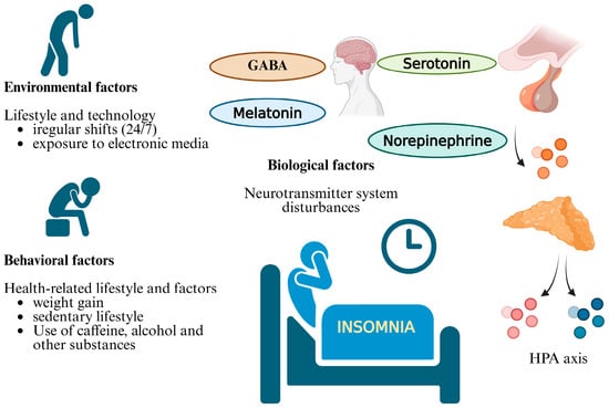
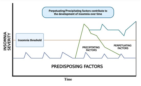
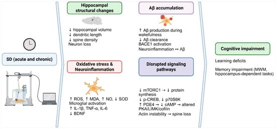
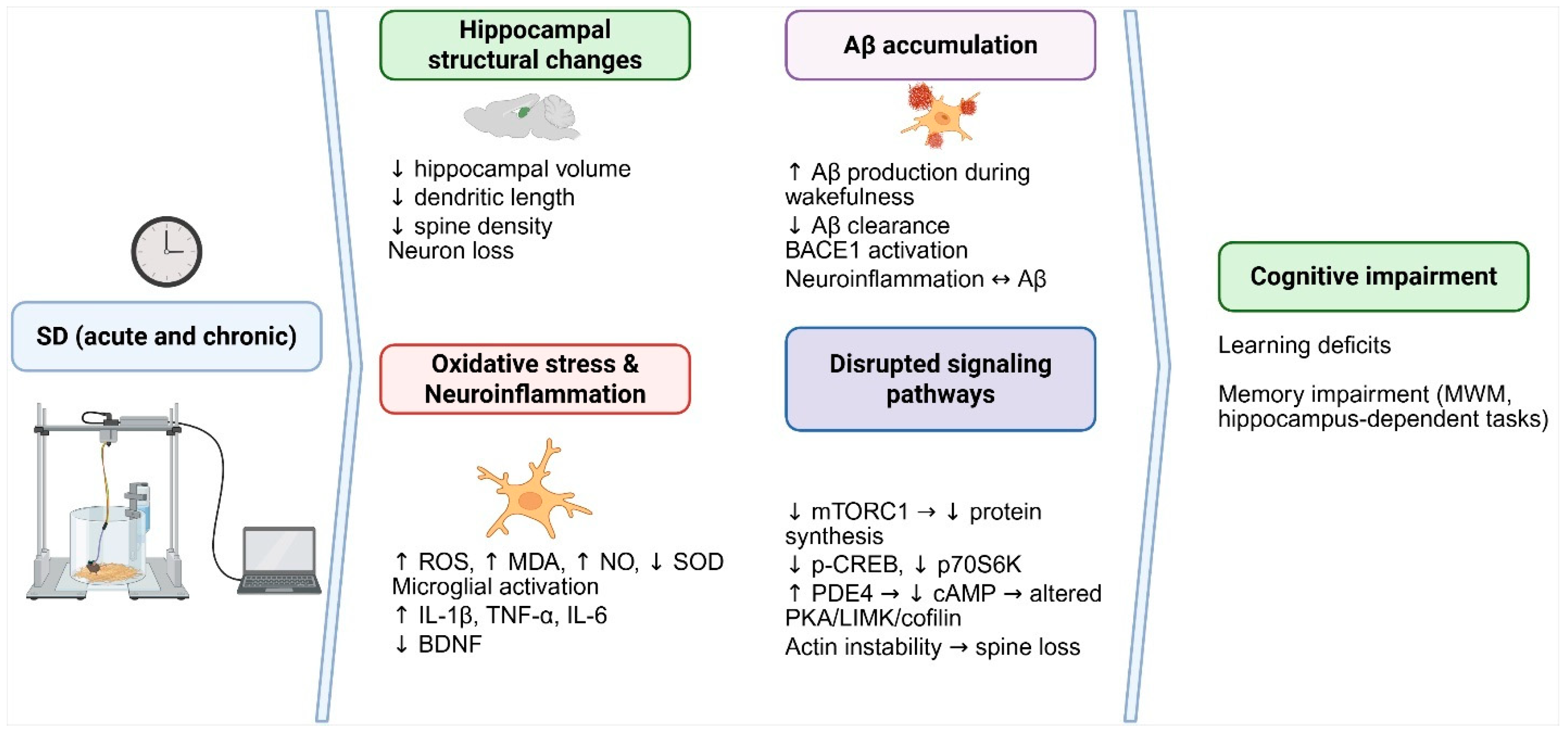

Disclaimer/Publisher’s Note: The statements, opinions and data contained in all publications are solely those of the individual author(s) and contributor(s) and not of MDPI and/or the editor(s). MDPI and/or the editor(s) disclaim responsibility for any injury to people or property resulting from any ideas, methods, instructions or products referred to in the content. |
© 2026 by the authors. Licensee MDPI, Basel, Switzerland. This article is an open access article distributed under the terms and conditions of the Creative Commons Attribution (CC BY) license.